


Abstract
Aim: The stromal cell-derived factor-1 (SDF-1)/C-X-C chemokine receptor type 4 (CXCR4) axis and Wingless and INT-1 (Wnt)/β-catenin pathway has been related to cancer progression. The aim of this study was to investigate the expression of CXCR4 and β-catenin in pancreatic cancer. Patients and Methods: A total of 48 pancreatic cancer samples and 8 normal pancreatic tissues were selected to detect CXCR4 and β-catenin expression by an immunohistological technique. Spearman and Chi-square analyses were used to study the relation between the protein expression and clinical characteristics. Survival analysis was evaluated by the Kaplan–Meier product limit method. Results: The proportions of CXCR4 and β-catenin expression on pancreatic cancer cells were significantly higher than in normal pancreas tissues. There was a significant difference in CXRC4 expression levels, lymph node metastasis and TNM stage. Clinical Significance was observed for β-catenin expression and lymph node metastasis; Kaplan-Meier curves suggested that clinical prognosis is poor for patients expressing CXCR4. Multivariate analysis showed that CXCR4 expression was an independent prognostic factor for pancreatic cancer. Conclusion: Both CXCR4 and β-catenin are abnormally expressed in pancreatic cancer. CXCR4 may be an important marker for pancreatic cancer progression.
Pancreatic cancer is the fourth leading cause of death from malignant cancers (1). At the time of diagnosis, more than 80% of patients with pancreatic cancer have locally advanced or metastatic lesions and fewer than 5% of patients survive five years after the diagnosis (2). The mechanism of metastasis remains unclear; however, molecular research has indicated an important relationship between cancer progression and chemokine receptors, particularly C-X-C chemokine receptor type 4 (CXCR4) (3-6). It has been demonstrated that stromal cell-derived factor-1 (SDF-1)/CXCR4 interaction is a critical molecular determinant for events such as embryo development, mediation of immune inflammatory reaction, and modulation of the hematopoietic system (7). Recently, the SDF-1/CXCR4 pathway has been proven to be involved in simulating the metastatic process of different neoplasms (8, 9). The chemokine receptor CXCR4, which is expressed in as many as 23 different tumor types, is a particularly exciting new target (10-14). Research potentially suggests a critical role of CXCR4 on cancer progression.
β-Catenin is a key factor in the canonical Wingless and INT (Wnt) pathway and plays critical roles in embryonic development, inflammation cancer proliferation and metastasis (15-17). It has potentially dual functions: cell adhesion and cell signaling. The deregulation of Wnt/β-catenin pathway leads to a high metastatic ability of cancer cells and influences cell adhesion, migration, and transcription through interactions with different protein complexes in the development of cancer (16, 18, 19). Deregulation of Wnt signaling is a well-established hallmark of certain types of human cancer.
In one study of neural progenitors, it seemed that SDF-1 was able to modulate β-catenin to influence the canonical Wnt pathway and finally influence cell differentiation (20). There are limited reports on the relation between the SDF-1/CXCR4 and the Wnt pathways in cancer progression. Our previous in vitro study showed that CXCR4 knockdown inhibited Wnt/β-catenin pathway activity and its target gene expression, which resulted in the decrease of pancreatic cancer cell growth, cell tumorigenesis and invasive ability (21). With this knowledge, we were able to gain insight into the relationship between SDF-1/CXCR4 and the Wnt/β-catenin pathway in the progression of pancreatic cancer (22). Therefore, we were eager to investigate in vivo the expression of CXCR4 and β-catenin in pancreatic ductal carcinoma, and analyze the relationship between the expression of these proteins and clinical prognosis.
Patients and Methods
Patients and tissue samples. The patients included in the study were those who underwent curative or palliative surgery for pancreatic cancer at the Department of Hepatobiliary Surgery, the First Affiliated Hospital of Medical College, Xi'an Jiaotong University, P.R. China, from August 2006 to August 2010. Forty-eight tissue samples of pancreatic ductal carcinoma were collected. Eight tissue samples of normal pancreatic tissue were obtained from patients operated on pancreatic trauma, and were verified by pathological techniques. All samples were fixed with 10% formalin, uniformly embedded with paraffin, and stained with hematoxylin-eosin (HE). The mean age of patients overall was 58.5±10.3 years, with a range of 34-76 years. Informed consent was obtained from the patients or their legal representatives before the study. The study was approved by the Institutional Ethics Committee of Xi'an Jiaotong University (No. 001003226). The clinicopathological features of patients according to the tumor-node-metastasis (TNM) stage classification of the International Union Against Cancer (UICC) are shown in Table I (23). Postoperative survival was defined as the time that elapsed from the surgery to cancer-related death. The median follow-up time was 16.2 months.
Cell line and culture conditions. Different grade human pancreatic cancer cell lines: poorly differentiated (Panc-1, Miapaca-2), moderately differentiated (BxPC-3, AsPC-1), and well-differentiated (PC-2, Capan-1) were stored at the Department of Hepatobiliary Surgery, First Affiliated Hospital of Xi'an Jiaotong University. The cells were cultured in Dulbecco's Modified Eagle's Medium (DMEM, Invitrogen Corporation, Carlsbad, CA. USA) supplemented with 10% fetal bovine serum (FBS, Invitrogen) and penicillin (100 units/ml), streptomycin (0.1 mg/ml) at 37°C in an environment of 95% air and 5% CO2.
Immunohistochemical staining. Four-micrometer tissue sections from each tissue sample on glass slides were deparaffinized three times in xylene for 8 min each, rehydrated three times in a graded series of ethanol, and washed with phosphate-buffered saline (PBS) (pH 7.4). The slides were treated with 3% H2O2 for 15 min to block endogenous peroxidase activity. These sections were heated in 0.01 M sodium citrate buffer to retrieve antigen activity and then cooled at room temperature. Primary antibodies to CXCR4 and β-catenin were obtained from eBioscience (San Diego, CA. USA) and Santa Cruz Biotechnology (Santa Cruz, CA, USA), respectively. The sections were first incubated with antibody to CXCR4 (1:50) and to β-catenin (1:200) at 4°C overnight, then incubated with biotin-labeled secondary antibody for 30 min, and finally incubated with streptavidin-peroxidase for 30 min at room temperature. The tissue sections were developed with diaminobenzidine (DAB), lightly counterstained with hematoxylin, and mounted. Negative controls were obtained by substituting the primary antibody with PBS. The evaluation of target protein was performed simultaneously by two pathologists who had no knowledge of the patients' clinicopathological features. All slides were observed under a microscope, and representative photographs were taken.
Evaluation of immunostaining and semiquantitative analysis. A refined scoring system was used for the evaluation of cytoplasmic staining results for CXCR4 and β-catenin. The immunoreactive score (IRS) was calculated according to a previous study by determining the product of the intensity of immunostaining (none=0, weak=1, moderate=2, strong=3) and the percentage of positively stained tumor cells (none=0, <10%=1, 10–50%=2, 51–80%=3, >80%=4) (24). By multiplying both these components, an expression score (0–12) was obtained. For the final statistical analysis, the IRS value of 0 was ranked as 0 (negative); IRS values 1–3, as 1 (weak expression); IRS values 4-8, as 2 (moderate expression); and IRS values 9-12, as 3 (strong expression).
Relationship between clinicopathological features and expression of C-X-C chemokine receptor type 4 (CXCR4) and β-catenin.
Quantitative real time polymerase chain reaction (QT-PCR) to detect mRNA expression. A total of 2×10⁵ pancreatic cancer cells were harvested with Trizol Reagent (Invitrogen) to extract total RNA. cDNA synthesis was conducted as followed with the SYBR® ExScript™ RT-PCR kit (Takara Biotechnology Co. Ltd., Dalian, P.R. China) according to the manufacturer's instructions: 500 ng total RNA was mixed with 2 μl of 5× ExScript™ RTase buffer, 0.5 μl of dNTP mixture, 0.5 μl of random hexamers, 0.25 μl of ExScript™ Rtase, and 0.25 μl of RNase Inhibitor in a total volume of 10 μl. The reactions were performed at 42°C for 12 min, followed by inactivation of the reverse transcriptase at 95°C for 2 min. The cDNA was stored at −20°C. QT-PCR was performed on an ABI PRISM® 7300 Sequence Detection System (Applied Biosystems, Foster City, CA, USA) with SYBR Green Master Mix. The final reaction volume was 25 μl and contained 12.5 μl 2× SYBR® Premix Ex Taq™, 1.0 μl of each primer (10 μM), 0.5 μl 50× ROX reference dye, and 1.0 μl cDNA. The cycling conditions were as follows: initial denaturation at 95°C for 10 min, followed by 40 cycles of 95°C for 15 s, and 60°C for 60 s. Each measurement was performed in triplicate, and no-template controls were included for each assay. After PCR, a dissociation curve analysis was conducted. Glyceraldehyde-3-phosphatedehydrogenase (GAPDH) was applied as the internal housekeeping gene. Relative gene expression was calculated using the 2−ΔΔCT method compared to GAPDH. Primers used for GAPDH were: F: AGCCACATCGCTCAGACAC, R: GCCC AATACGACCAAATCC; for CXCR4 were: F:CTGTG AGCAGAGGGTCCAG, R: ATGAATGTCCACCTCGC TT; and for β-catenin were: F: GCTTTCAGTTGAGCTGACCA, R: AAGTCC AAGATCAGCAGTCTCA.

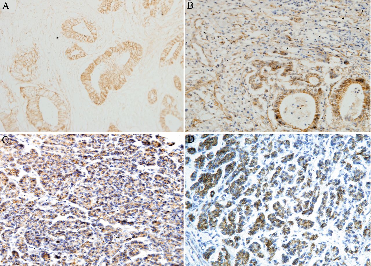
Immunohistochemical analysis showing different C-X-C chemokine receptor type 4 (CXCR4) expression levels in non-malignant pancreatic tissues (A) and pancreatic cancer tissues (B to D). A: Negative expression; B: weak cytoplasmic expression; C. moderate cytoplasmic expression; D. strong cytoplasmic expression (original magnification ×200).
Statistical analysis. All statistical tests were two-sided, and a probability value of <0.05 was considered as statistically significant. To evaluate the association between ordinal data of CXCR4 expression and β-catenin expression, the Spearman correlation coefficient was calculated. Correlation between the target protein and clinicopathological features was assessed using the Chi-square test. Survival was evaluated by the Kaplan–Meier product limit method and compared between groups using the log-rank test. Multivariate survival analysis for independent prognostic factors was performed by a Cox proportional hazards model that included significant univariate variables. A value of p<0.05 was considered statistically significant.
Results
CXCR4 and β-catenin are abnormally expressed in pancreatic ductal carcinoma. An immunohistological technique was applied for pancreatic ductal carcinoma and non-malignant pancreatic tissue samples. CXCR4 expression was not detected in the normal pancreatic tissues but was detected in the cytoplasm of most pancreatic ductal carcinomas. β-Catenin was predominantly expressed in the cytomembrane of normal pancreas cells. However, in pancreatic cancer samples, down-regulation of membrane expression and enhancement of cytoplasmic expression were observed for β-catenin. Positively stained tumor cells exhibited a diffuse cytoplasmic staining. Representative staining results are shown in Figures 1 and 2. Abnormal staining of CXCR4 and β-catenin was found in 85.4% (41 out of 48) and 75.0% (36 out of 48) cancer samples, respectively. To investigate the correlation of the two proteins, we analyzed the coexpression of CXCR4 and β-catenin on the same specimen. Cytoplasmic coexpression of CXCR4 and β-catenin was observed in 70.8% (34 out of 48) of cancer tissue samples, and negative expression of both these proteins was observed in 10.4% (5 out of 48) of samples. The Spearman correlation coefficient test revealed a significantly positive correlation between CXCR4 and β-catenin in the investigated tumor samples (rs=0.443, p=0.002).
CXCR4 and β-catenin are abnormally expressed in pancreatic cancer cell lines. In the current study, six pancreatic cancer cell lines were divided into three groups according to their grade of differentiation. As Figure 3 shows, CXCR4 was significantly more highly expressed in the poorly-differentiated cell lines Miapaca-2 and Panc-1 than moderately-differentiated cell lines BxPC-3 and AsPC-1 (p <0.05). The expression of CXCR4 was lowest in the well-differentiated cell lines PC-2 and Capan-1. It seems that the expression of CXCR4 was down-regulated according to the malignancy of the pancreatic cancer cells. Similar results were also observed for the expression of β-catenin in the pancreatic cancer cell lines (Figure 4). The expression of β-catenin mRNA in the poorly differentiated cell lines was four fold higher than in the well-differentiated cell lines.

Different expression levels of β-catenin in non-malignant pancreatic tissues (A) and pancreatic cancer tissues (B to D). A: Membrane expression; B: weak cytoplasmic expression; C: moderate cytoplasmic expression; D: strong cytoplasmic expression (original magnification ×200).
CXCR4 and β-catenin expression in patients with pancreatic cancer: Correlation with clinicopathological features. As shown in Table I, the expression of both CXCR4 and β-catenin in the investigated tumor samples had no significant correlation with the clinicopathological features of the patients (e.g. gender, age, tumor site, and histological grade). Based on the analysis of association between different protein staining levels and clinicopathological features, we classified the expression into four levels: none, weak, moderate, and strong. We calculated the IRS score for both proteins. Statistical analysis revealed no association of the expression of CXCR4 or β-catenin with gender, age, tumor site, and histological grade. However, as Table II shows, a significant difference was observed for different CXCR4 expression levels: IRS increasing with lymph node metastasis (p=0.012) and with increasing TNM stage (p=0.005). We also found that the IRS significantly increased for β-catenin expression with lymph node metastasis (p=0.047).
CXCR4 and β-catenin expression in patients with pancreatic cancer: Correlation with overall survival. Two patients died during the follow-up period. Most of the study patients died after a median follow-up period of 16.2 months. The 1-year and 2-year survival rates were 20.8% and 4.2%, respectively. For CXCR4 expression alone, the median survival time of the CXCR4-positive and CXCR4-negative groups was 5.5 and 13.5 months, respectively. The pattern of the Kaplan–Meier estimated survival curves suggests that prognosis was poor in patients expressing CXCR4 (Figure 5). The median survival time of the β-catenin positive and β-catenin-negative groups was 5.6 and 8.0 months, respectively. No significant difference was observed in the median survival time for β-catenin expression (Figure 6). It seems that abnormal expression of β-catenin did not determine the clinical survival. The log-rank test revealed that increased CXCR4 expression was a significant predictor of overall survival (p=0.002), while β-catenin was not (p=0.17). Multivariate analysis by the Cox proportional hazard model indicated a significant correlation between clinical prognosis and CXCR4-positive expression, late TNM stage (III+IV), and presence of lymph node metastasis (p=0.014, 0.016, and 0.014, respectively). However, β-catenin expression alone was observed to have no association with patients' survival (p>0.05).

C-X-C chemokine receptor type 4 (CXCR4) mRNA expression in different pancreatic cancer cell lines. CXCR4 was significantly more highly expressed in the poorly differentiated cell lines Miapaca-2 and Panc-1 than moderately differentiated cell lines BxPC-3 and AsPC-1 (p<0.05). The expression of CXCR4 was lowest in the well-differentiated cell lines PC-2 and Capan-1.

β-Catenin mRNA expression in different pancreatic cancer cell lines. The expression of β-catenin mRNA level in the poorly and low differentiated cell lines was four-fold higher than in the well-differentiated cell lines.
Association between clinicopathological features and different levels of C-X-C chemokine receptor type 4 (CXCR4) and β-catenin expression.
Relationship between various factors and clinical prognosis.
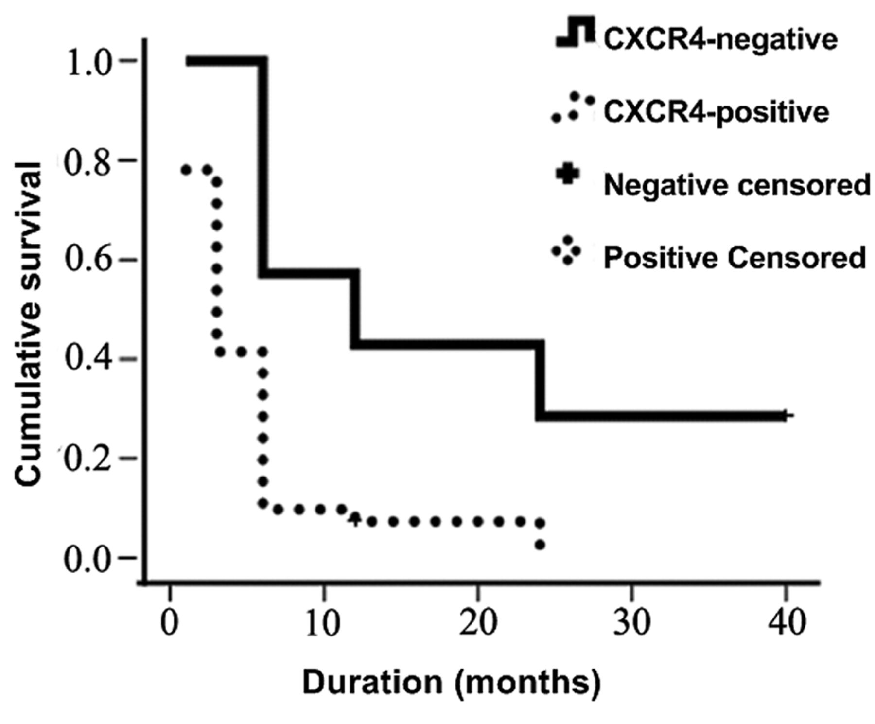
Kaplan–Meier product limit estimate of disease-free survival of patients with pancreatic cancer based on C-X-C chemokine receptor type 4 (CXCR4) expression.
Discussion
Pancreatic ductal carcinoma is a highly malignant tumor, always accompanied by cancer metastasis. Metastasis of malignant tumor results from a series of complex processes that depend on multiple factors. Thus far, the obvious effects of chemokines on cancer metastasis have received considerable attention (4). Chemokine receptors have been demonstrated to play a critical role in the homing of metastatic tumor cells. CXCR4 is a major chemokine receptor and is reported to be expressed in most solid types of cancer such as of the breast, prostate, ovary, and colon. Additionally, it was observed that CXCR4 has a significant correlation with organ-specific metastasis in some types of cancer. Recent studies have shown that organ-specific metastasis could be inhibited by neutralizing CXCR4 antibody and siRNA technique in vivo and in vitro (3, 25, 26). Therefore, CXCR4 appears to be a prominent target gene for the therapy of tumor metastasis.
A previous study showed that inhibition of CXCR4 effectively blocked pancreatic cancer progression in vitro through the canonical Wnt pathway (21). In the present study, we focused on CXCR4 expression in pancreatic cancer. We observed that CXCR4 expression in pancreatic ductal carcinoma was significantly higher than that in normal pancreas. This finding is in agreement with the findings of Koshiba et al. who showed that CXCR4 was expressed in most pancreatic cancer tissues (27). Classification of CXCR4 expression into different stages revealed a strong relationship between CXCR4 and clinicopathological features such as lymph node metastasis and late TNM stage. Therefore, it seems that CXCR4 is possibly a major protein that influences the malignant behavior of pancreatic cancer. Other investigators have reported that CXCR4 expression did not correlate with clinicopathological features, including metastasis and TNM stage, for some types of tumors (28, 29). Thus, there is a discrepancy regarding the clinical significance of CXCR4 in cancer. Although we cannot explain this discrepancy at present, we speculate that CXCR4 expression in cancer cells is an important factor in cancer progression, at least in patients with pancreatic cancer.

Kaplan-Meier product limit estimate of disease-free survival of patients with pancreatic cancer based on β-catenin expression.
Several previous studies have suggested that aberrant expression of β-catenin protein is associated with tumor progression, metastasis, and poor patient prognosis in many types of cancer, and this may be attributable to its role in cell adhesion and influence on the epithelial-mesenchymal transition (EMT) process (30-33). In this study, membrane expression of β-catenin was down-regulated and its cytoplasmic expression was up-regulated in late tumor stages. It is conceivable that altered β-catenin expression and location contribute to pancreatic cancer progression because β-catenin is involved in cell proliferation, cell adhesion, and cell death. Our data indicated that abnormal β-catenin expression was related to lymph node metastasis, but our data show that there was no significant difference in the median survival time by β-catenin expression. We consider that multiple factors, including β-catenin, influence clinical survival, but β-catenin doe not determine survival.
Abnormal expression of CXCR4 and β-catenin was simultaneously observed in 70.8% of samples. Our data also showed that the expression of CXCR4 and β-catenin was higher in poorly-differentiated cell lines than in moderately-differentiated and well-differentiated cell lines. That is to say, the expression of CXCR4 and β-catenin changed in parallel with the progression of pancreatic cancer. This suggests that these two protein biomarkers may be inter-related. Moreover, the Spearman correlation coefficient analysis revealed a significant relationship between CXCR4 and β-catenin expression. SDF-1 is known to stimulate β-catenin through the CXCR4 receptor. β-Catenin is not easily up-regulated and altered when CXCR4 is inhibited (20). Our in vitro study further proved the relationship between the SDF-1/CXCR4 axis and the Wnt/β-catenin pathway in the progression of pancreatic cancer.
For the early detection of pancreatic cancer, there are currently no available effective molecular markers. Therefore, research has been focused on the early detection of pancreatic ductal carcinoma. The strong expression of CXCR4 was related to advanced disease. Our Kaplan–Meier estimated curves revealed obvious differences between the CXCR4-positive and CXCR4-negative groups with respect to clinical prognosis. Statistical analysis also revealed that CXCR4 expression was a predictor of overall survival. Thus, adjusting for the TNM stage and metastasis, CXCR4 expression could predict the clinical prognosis of pancreatic cancer. It seems that CXCR4 could be a promising molecular marker for pancreatic cancer. Although β-catenin may influence the progression of pancreatic cancer, from our data it does not appear to predict its prognosis.
Acknowledgements
We thank Dr Le Zhao for her expert technical assistance. We would like to give special thanks to Dr. Li Li and Dr. Guodong Zhu for their critical discussion. This study was supported partly by the National Natural Science Foundation of China (no. 30700817, 81172195); and the Fundamental Research Funds for the Central Universities at Xi'an Jiaotong University (2011-6).
- Received June 16, 2013.
- Revision received July 2, 2013.
- Accepted July 4, 2013.
- Copyright© 2013 International Institute of Anticancer Research (Dr. John G. Delinassios), All rights reserved
References
In this issue


Jump to section
Related Articles
Cited By...
- {beta}-catenin plus PROX1 immunostaining stratifies disease progression and patient survival in neoadjuvant-treated pancreatic cancer
- Co-targeting of CXCR4 and hedgehog pathways disrupts tumor-stromal crosstalk and improves chemotherapeutic efficacy in pancreatic cancer
- First Experience with Chemokine Receptor CXCR4-Targeted PET Imaging of Patients with Solid Cancers