


Abstract
Background: We examined the levels of long non-coding RNAs (lncRNAs) in the plasma of patients with gastric cancer to assess their clinical significance for diseases diagnosing and monitoring. Materials and Methods: We investigated the stability of plasma lncRNAs, and then confirmed the appropriateness of the lncRNA assay with a pre-amplification method. The levels of plasma lncRNAs, H19, HOX antisense intergenic RNA (HOTAIR), and metastasis associated lung adenocarcinoma transcript-1 (MALAT1), were then analyzed in patients with gastric cancer (GC) and healthy controls. Results: Plasma lncRNAs exhibited minimal gradual instability only under several severe conditions. Analysis showed that samples with pre-amplification had a higher level of linearity in the reverse transcription polymerase chain reaction (RT-PCR) assay than those without pre-amplification. Plasma H19 levels were significantly higher in patients than in healthy controls. Plasma H19 levels were significantly reduced in postoperative samples. Conclusion: Circulating lncRNAs can be detectable in plasma, and the detection of circulating lncRNAs may provide new complementary tumor markers for gastric cancer.
Gastric cancer is the second leading cause of cancer-related death in the world (1). The prognosis of advanced gastric cancer remains dismal in spite of recent improvements in therapeutic methods such as extended radical operations and perioperative chemotherapies (2). Thus, in order to improve the cure rates for patients with gastric cancer, primary tumors must be detected at an early stage and recurrent disease must be diagnosed when it is still minimal or clinically occult (3).
Biomarkers have attracted attention for early detection, prediction of prognosis, and chemotherapeutic sensitivity in patients with various types of cancer (4). Among the various approaches currently used, blood-based testing is an ideal method for biomarkers in cancer care due to its ease and lower invasiveness. However, conventional serum biomarkers, such as carcinoembryonic antigen (CEA) and carbohydrate antigen (CA) 19-9, lack sufficient sensitivity and specificity. Several studies have demonstrated that tumor-specific or tumor-associated alterations in circulating nucleic acids may serve as new complementary tumor markers for gastric cancer. In recent years, cell-free RNA has been stably detected in the plasma and serum similarly to other molecules (5-7).
Over the past few decades, advances in genome-wide analyses have identified that almost all of the human genome is transcribed and produces large numbers of long non-coding RNAs (lncRNAs) (8-10). Recently, several studies have demonstrated that some lncRNAs are involved in the development of various types of cancer (11, 12). The levels of some lncRNAs, such as H19 and colon cancer-associated transcript-1 (CCAT1), were reportedly markedly increased in gastric cancer, and studies suggested important roles for lncRNAs in the molecular etiology of gastric cancer (13, 14). These findings prompted us to examine the detectability of lncRNAs in the blood samples of patients with gastric cancer.
In this study, we investigated the levels of circulating lncRNAs in plasma samples from both patients with gastric cancer and from controls, and compared the relationships between the results obtained and clinicopathological findings to assess the diagnostic value of these biomarkers in patients with gastric cancer.
Materials and Methods
Ethics statement. Ethical approval was granted by the Faculty of Science Ethics Committee at the Kyoto Prefectural University of Medicine (RBMR-C-179) and the study was conducted in accordance with the principles of the Declaration of Helsinki. Written informed consent was obtained from all patients and healthy volunteers.
Patients and samples. A total of 43 pre-operative plasma samples were collected from patients with gastric cancer who underwent gastrectomy at the Kyoto Prefectural University of Medicine between 2008 and 2012. Of these patients, 20 post-operative plasma samples were also collected one month after surgery. Thirty-three control samples were collected from healthy volunteers without cancerous diseases. Relevant clinical and survival data were available for all patients. Formalin-fixed paraffin-embedded tumor samples were also examined in patients whose plasma H19 levels were determined. Resected gastric cancer specimens were fixed in buffered formalin and embedded in paraffin for pathological examination using standard methods. Macroscopic and microscopic classifications of tumors were based on the International Union against Cancer/Tumor, Node, Metastasis (UICC/TMN) staging system (15) and Japanese classification of gastric carcinoma: 3rd edition (16).
Stock of plasma samples. Blood samples were subjected to the isolation of cell-free nucleic acids immediately after collection using a 3-spin protocol (350 rcf×g for 30 min, 700 rcf ×g for 5 min, 1600 rcf×g for 5 min) to prevent contamination by cellular nucleic acids. Plasma samples were then stored at −80°C until further analyses.
RNA extraction. Total RNA was extracted from cultured cells and 400 μl of plasma using a mirVana PARIS Kit (Ambion, Austin, TX, USA), and was eluted into 100 μl of pre-heated (95°C) Elution Solution according to the manufacturer's instructions. The total RNA of tissue samples was extracted from four slices of 15 μm-thick formalin-fixed paraffin-embedded tissue (with a total thickness of 60 μm) using the RecoverAll Total Nucleic Acid Isolation Kit (Ambion), and was finally eluted into 60 μl of Elution Solution according to the manufacturer's instructions. RNA samples were stored at −80°C until further processing.
Protocol for the detection of lncRNAs [H19, HOX antisense intergenic RNA (HOTAIR), and metastasis associated lung adenocarcinoma transcript 1 (MALAT1)]. The reverse transcription reaction of 9 μl of total RNAs extracted from 400 μl of plasma was carried out with the High-Capacity RNA-to-cDNA Kit (Applied Biosystems, Foster City, CA, USA). Reacted cDNAs were then pre-amplified using the TaqMan PreAmp Master Mix Kit (Applied Biosystems), according to the manufacturer's instructions. In this process, each lncRNA-specific primer of the human TaqMan Gene Expression Assay Kit (Applied Biosystems) was used. The levels of lncRNAs were quantified in duplicate by quantitative real time-polymerase chain reaction (PCR) using the human TaqMan Gene Expression Assay Kit (Applied Biosystems) following the manufacturer's protocol. In brief, quantitative PCR analyses were performed using the Step One Plus Real-time PCR system (Applied Biosystems) and cycle threshold (Ct) values were calculated with the Step One Software version 2.2.2 (Applied Biosystems). The levels of lncRNAs in plasma were calculated using the ΔΔCt method relative to the plasma level of lncRNAs in one patient with gastric cancer (GC498) because there has been no consensus as yet, concerning stable and suitable internal controls for non-coding RNAs in plasma samples. The expression of H19 from tissue samples was also normalized using ΔΔCt method relative to actin β (ACTB). The change in gene expression was calculated with the equation 2−ΔΔCt.
Cell culture. The human stomach carcinoma cell line HGC27 (RCB0500) was purchased from RIKEN Bio Resource Center (Tokyo, Japan), and maintained in DMEM (Nacalai Tesque, Kyoto, Japan) supplemented with 10% fetal bovine serum (Trace Scientific, Melbourne, Vic., Australia), 100 U/mL penicillin, and 100 μg/mL streptomycin. The flask was kept in a humidified incubator at 37°C under 5.0% CO2 in air.
Statistical analysis. The paired t-test was used to evaluate differences in the levels of RNA in the GC cell line and plasma with/without pre-amplification. The Wilcoxon test was used to compare paired plasma samples obtained pre- and post-surgery, and the Mann-Whitney test was used to compare for differences in the levels of plasma RNA between the cancer group and healthy group. A p-value less than 0.05 were considered significant. Receiver operating characteristic (ROC) curves and the area under the ROC curve (AUC) were used to assess the feasibility of using plasma H19 as a diagnostic tool for the detection of gastric cancer.
Results
Stability of lncRNAs in human plasma. To investigate the stability of lncRNAs in plasma, we firstly evaluated changes in the levels of H19 in plasma under oppressive conditions. Plasma H19 exhibited minimal gradual instability only in several severe conditions, such as the incubation of plasma at room temperature for up to 24 h, three cycles of freeze-thawing processes, and also incubation at 45°C for 24 h (Figure 1A-C, respectively).
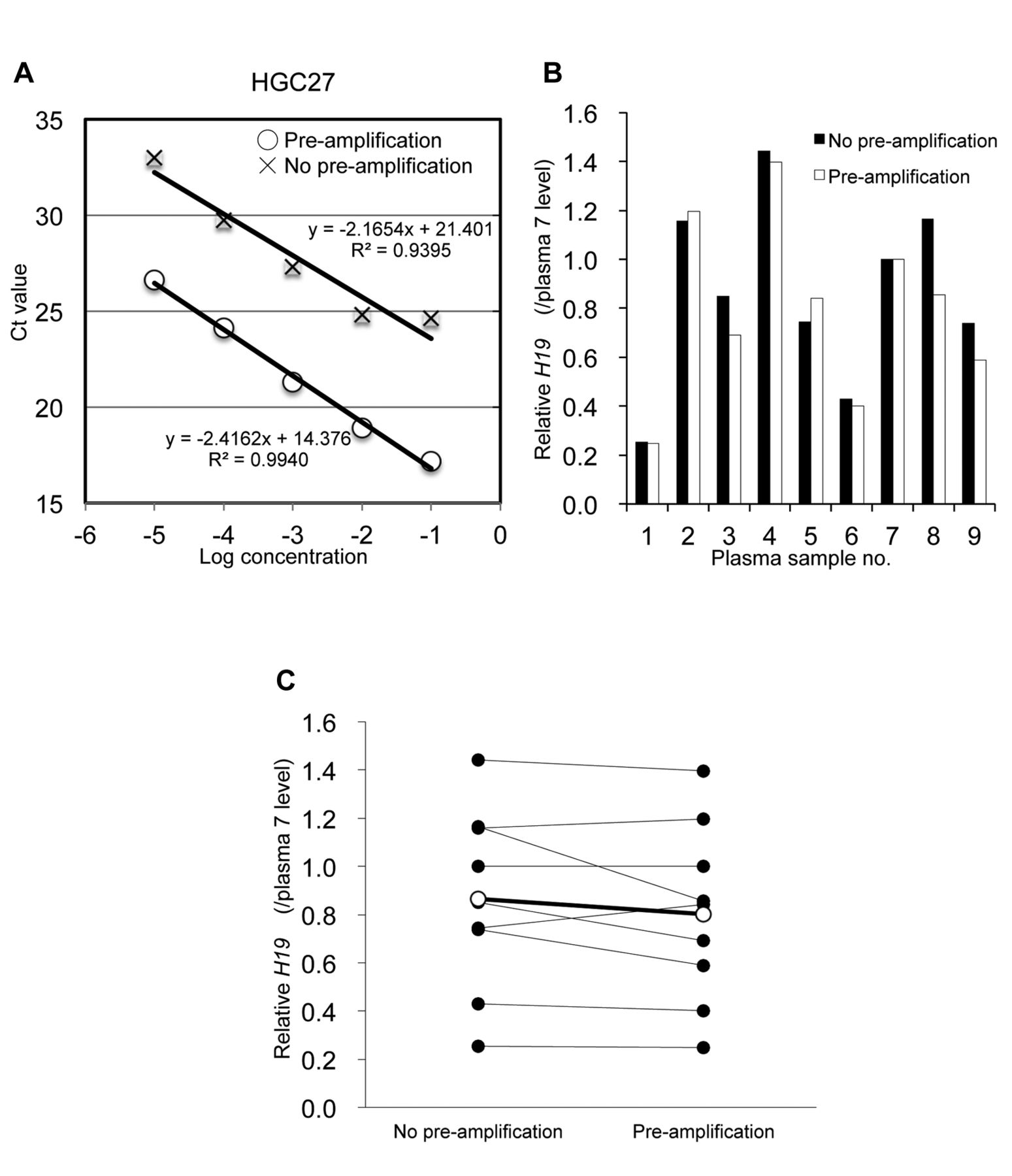
Applicability of the pre-amplification assay. To evaluate the appropriateness of the pre-amplification method, which was encouraged by the manufacturer's protocol, we conducted amplification of H19 by the real-time RT-PCR assay of a 10-fold serial dilution using the total RNA of a gastric cancer cell line (HGC27), and also compared the levels of H19 in plasma samples with and without pre-amplification (Figure 2A and B). Analysis showed that samples which underwent pre-amplification had a higher level of linearity in the RT-PCR assay than those without pre-amplification (R2=0.9940 vs. 0.9395, respectively), which indicates the superior quantitative performance of the pre-amplification assay. Moreover, the relative levels of circulating H19 were approximately the same with and without pre-amplification in each plasma sample from patients with gastric cancer (Figure 2C).
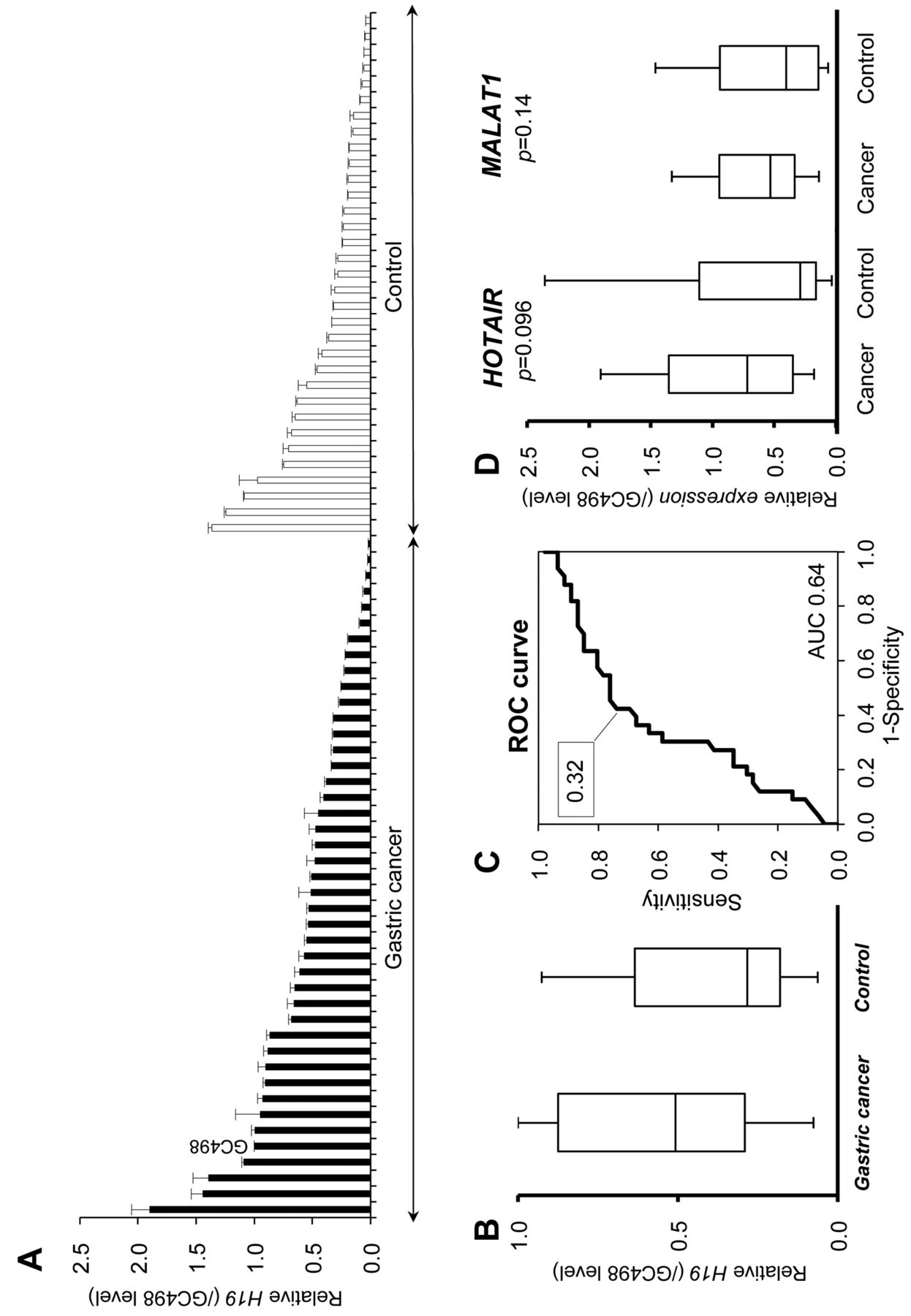
Comparison of the levels of lncRNAs in plasma between healthy controls and patients with gastric cancer. Plasma H19 levels were significantly higher in patients than in healthy controls (p=0.029, Figure 3A and B); however, plasma HOTAIR and MALAT1 levels were not (Figure 3D). The value of the area under the ROC curve was 0.64 for the plasma H19 assay (Figure 3C). In this model, optimal cut-off points were indicated at 0.32 (sensitivity 74% and specificity 58%). The clinicopathological features of these patients are shown in Table I; however, there was no correlation between plasma H19 levels and clinicopathological factors.
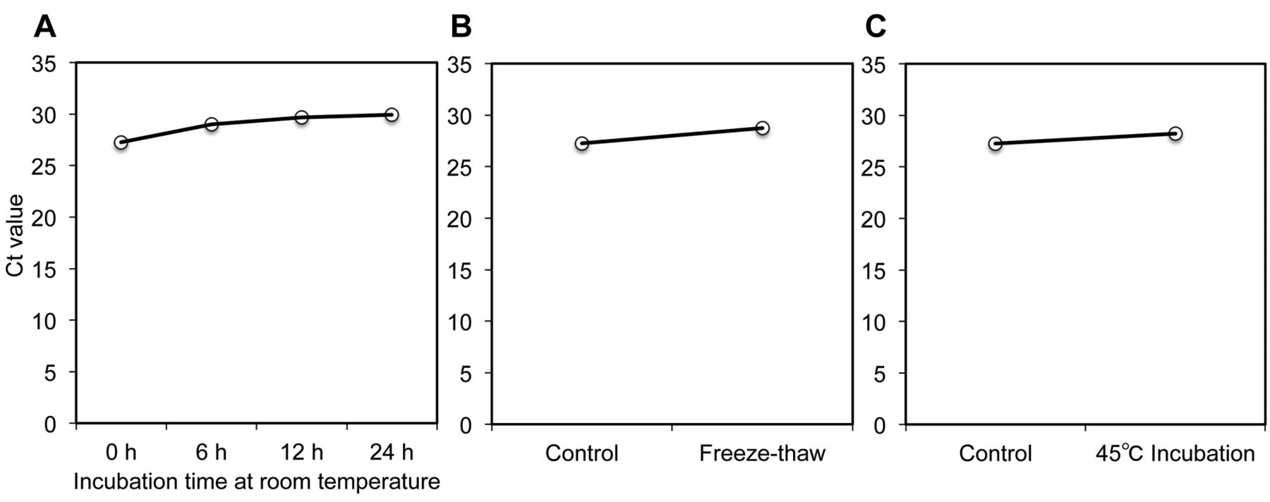
Stability of H19 in human plasma under oppressive conditions. Plasma H19 exhibited minimal gradual instability with the incubation of plasma at room temperature (A), freeze-thawing processes (B), and incubation at 45°C (C). Levels are presented as the Ct value.
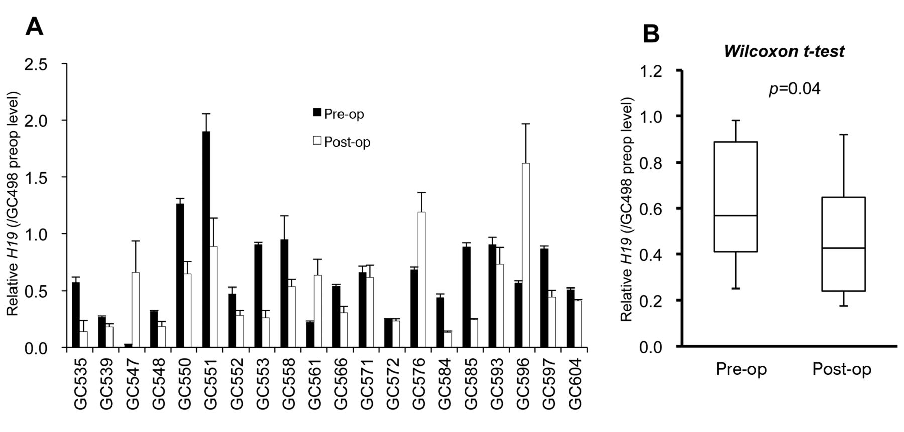
Comparison of H19 levels in pre- and postoperative paired plasma samples, and in paired cancerous and non-cancerous tissues. H19 levels were significantly lower in postoperative plasma than in pre-operative plasma in 16 cases (16/20; p=0.047, Wilcoxon t-test, Figure 4A and B). However, there was no significant difference in H19 expression between cancerous tissues and paired non-cancerous tissues using paraffin-embedded tissues (p=0.14, Wilcoxon t-test, Figure 5).
Discussion
Tumor development and the progression of gastric cancer are multistep processes involving numerous genetic and epigenetic alterations. Only several oncogenes and tumor suppressor genes coding for proteins were previously considered to be involved in gastric carcinogenesis. However, recent advances in sequencing technologies have revealed that the majority of transcriptional units are non-coding RNAs, which also play important roles as oncogenes or tumor suppressor genes in various types of cancer (17-20). Recently, some lncRNAs, as well as microRNAs, have been reported to play important roles in carcinogenesis (13, 14, 21, 22). Detailed investigations have shown that lncRNAs are involved in fundamental cellular processes, such as RNA processing, gene regulation, chromatin modification, gene transcription, and post-transcriptional gene regulation on the basis of RNA sequence complementary interactions (23, 24). Although the functions of all lncRNAs have not been fully-clarified, the molecular mechanisms of a few lncRNAs, such as MALAT1, Highly Up-regulated in Liver Cancer (HULC), H19 and HOTAIR have been elucidated to some degree (22, 23-28). Moreover, the altered expressions of lncRNAs have been increasingly reported in a variety of cancer types, which indicates a possible connection between lncRNAs and carcinogenesis (29).
The H19 gene encodes a 2.3-kb lncRNA and is a paternally imprinted gene located at 11p15.5. H19 is expressed during embryonic development, and is down-regulated in most tissues after birth (30). Several recent studies have demonstrated that H19 is strongly expressed in cancer, such as esophagus, colorectal, liver, endometrial, cervix, and bladder cancer, in which H19 has an oncogenic function (31-35). Yang et al. demonstrated that levels of the lncRNA H19 were markedly higher in gastric cancer cell lines and also cancer tissues than in normal controls (13). They also confirmed an increase in cell proliferation by ectopic H19 expression and the induction of apoptosis by H19 siRNA treatment, and suggested an important role for H19 in the molecular etiology of gastric cancer.
Recent reports have demonstrated that microRNAs are extremely stable in plasma and have diagnostic potential in the early detection of several malignancies (7, 36-42). Therefore, it is possible that other types of non-coding RNA, such as lncRNAs, are also stable in plasma and have diagnostic potential in cancer management. However, only a few studies have shown that lncRNAs can be detected in the body fluids of patients with cancer (21, 43). Therefore, in the present study, we firstly confirmed if lncRNAs were detectable in plasma, and then investigated the stability of circulating lncRNAs under several severe conditions. Our findings clearly demonstrated that lncRNAs were relatively stable in plasma samples, and were protected by some mechanism(s) from the severe conditions tested. In fact, we have already confirmed that plasma exosomes contain a certain level of extracellular lncRNAs (data not shown). The mechanisms accounting for the stability of plasma lncRNAs are not well-understood; they may be protected by exosome encapsulation and also complex formation with proteins, such as plasma microRNAs (44, 45).

Applicability of the pre-amplification assay. A: Standard curve of H19 by the real-time RT-PCR assay of a 10-fold serial dilution of total RNA in HGC27 (gastric cancer cell line). Linearity was superior in samples with pre-amplification than in those without pre-amplification. B: Plasma H19 levels are presented as a ratio relative to that of plasma 7. The levels of plasma H19 were similar regardless of pre-amplification. C: There was no significant difference in H19 levels with and without pre-amplification (p=0.084).
A pre-amplification method is recommended for the lncRNAs assay by the manufacturer's protocol because of low concentrations. Therefore, before the analysis of clinical samples, we compared the linearity between the logarithms of the level of input lncRNAs and the cycle threshold value on real-time PCR with and without pre-amplification, and confirmed that methods with pre-amplification provide high quantitative performance even for low concentrations, and are an appropriate for plasma assays.

Comparison of plasma lncRNAs between healthy controls and patients with gastric cancer. A: Circulating plasma H19 was detectable and amplified in all samples from 43 patients and 34 healthy controls. The results are presented as a ratio relative to that of the gastric cancer sample 498. Each column shows the mean for duplicate experiments; bars=S.D. B: Plasma H19 levels were significantly higher in patients than in healthy controls (p=0.029, Mann–Whitney U-test). The upper and lower limits of the boxes and the line inside the boxes indicate the 75th and 25th percentiles and the median, respectively. C: Receiver-operating characteristic curve analyses in the plasma H19 assay for detecting gastric cancer patients (area under the curve=0.64). D: The levels of plasma HOX antisense intergenic RNA (HOTAIR) and metastasis associated lung adenocarcinoma transcript 1 (MALAT1). There were no significant differences in these levels between patients and healthy controls (p=0.096 and 0.14, respectively, Mann–Whitney U-test).
Correlation between H19 expression level and clinicopathologic factors.
Based on these findings, we compared the levels of circulating lncRNAs in plasma samples from both patients with gastric cancer and controls. Because there has never been a consensus about a universal internal control for the plasma lncRNAs assay, in this study we utilized the absolute concentration method for measuring plasma lncRNAs. H19 levels were significantly higher in patients than in controls, and the area under the ROC was 0.64. However, there was no significant difference in the levels of HOTAIR and MALAT1 between patients and controls. The value of AUC was not so high; however, adding analyses for other related lncRNAs may increase the sensitivity and the specificity.
We then measured the circulating H19 in paired-plasma before and one-month after the surgical removal of tumors in order to confirm the tumor release of H19. The results showed that the levels of H19 were significantly reduced postoperatively in patients with high preoperative plasma H19 levels, which may reflect the possible release of plasma lncRNAs from primary gastric tumors. However, there was no significant correlation between the expression of H19 in plasma and primary tumor tissues in the present study. These discrepancies remain to be clarified; however, a possible explanation for this finding may be the heterogeneity of primary tumors or the effects of formalin-fixation and paraffin-embedding on the stability of lncRNAs.
Plasma lncRNA assays may have several potential clinical applications in cancer management: i) screening and early diagnosis, ii) evaluation of surgical or non-surgical therapeutic efficiency, and iii) monitoring for recurrence during the follow-up period. We analyzed only a few cancer-related lncRNAs in this study; however, adding analyses for other related lncRNAs may increase clinical utility. A more intriguing possibility is that circulating lncRNAs may also have some function as an inter-cellular communication tool, such as extracellular miRNAs. However, there are still some limitations that need to be addressed before the measurement of circulating lncRNAs can be utilized in the clinical setting. There has been no consensus regarding which extraction and detection method is suitable for plasma lncRNAs, nor as to what molecule is the most appropriate for use as an endogenous control. Further studies are needed to verify the clinical usefulness of circulating lncRNA assays for each potential application, and also to elucidate the functional aspects of extracellular lncRNAs.

Comparison of plasma H19 between pre- and postoperative samples from patients with gastric cancer. The results are presented as a ratio relative to that of the preoperative gastric cancer sample 498 shown in Figure 3. A: Plasma H19 levels were higher in preoperative samples than in postoperative samples in 16/20 patients. Each column is the mean for duplicate experiments; bars=S.D. B: Box plots of pre- and postoperative plasma H19 concentrations. The levels were significantly reduced in postoperative plasma samples (p=0.047, Wilcoxon t-test). The upper and lower limits of the boxes and the lines inside the boxes indicate the 75th and 25th percentiles and the median, respectively. The upper and lower horizontal bars denote the 90th and 10th percentiles, respectively.

Comparison of H19 expression levels in gastric cancer tissues and non-cancerous tissues. A: Expression levels are presented as a ratio relative to that of non cancerous tissue from gastric cancer case 272. B: The box plots of H19 in cancerous tissue and non-cancerous tissue. There was no significant difference in the levels of H19 between the tissues (p=0.14).
In conclusion, the detection of circulating lncRNAs may serve as a new complementary marker for gastric cancer. This assay may be helpful in detecting patients with primary gastric cancer at an early stage and also clinically occult recurrence during the follow-up period after gastrectomy. Further prospective clinical trials using a variety of plasma lncRNAs should be carried out to define the usefulness of the assay for each potential application.
Footnotes
-
↵* These Authors contributed equally to this work.
- Received May 10, 2013.
- Revision received June 13, 2013.
- Accepted June 14, 2013.
- Copyright© 2013 International Institute of Anticancer Research (Dr. John G. Delinassios), All rights reserved
References
In this issue


Jump to section
Related Articles
Cited By...
- Exosomal Functional Cargoes from Liquid Biopsy of Gastric Cancer: A Systematic Review of Studies With Potential Clinical Relevance
- H19 in Endocrine System Tumours
- Inhibition of E-cadherin expression by lnc-RNA H19 to facilitate bladder cancer metastasis
- Circulating Noncoding RNAs as Biomarkers of Cardiovascular Disease and Injury
- Plasma long non-coding RNA, CoroMarker, a novel biomarker for diagnosis of coronary artery disease
- Long Noncoding RNA in Digestive Tract Cancers: Function, Mechanism, and Potential Biomarker
- Identification and Initial Functional Characterization of a Human Vascular Cell-Enriched Long Noncoding RNA