


Abstract
Aim: Advanced renal cancer has still a very poor prognosis. We combined wingless-related integration site (WNT) inhibitors with a bi-functional peptide, as previous research has proven their individual efficacy in cancer therapy. Each targets cancer cells differently. We wanted to determine whether they have an additive effect. Materials and Methods: Our bi-functional peptide consists of a target domain (LTVSPWY) and a lytic domain (KLAKLAK)2. We used three WNT inhibitors: Ethacrinic acid, ciclopirox olamine, piroctone olamine and combined each with the bi-functional peptide. They were tested on three different renal cancer cell lines using 3-(4,5-dimethylthiazol-2-yl)-2,5-diphenyltetrazolium-bromide (MTT) assay. Results: We demonstrated a synergistic effect of WNT inhibitors with the bi-functional peptide. The vitality of cancer cells was reduced significantly (p<0.05), while healthy cells were mostly unaffected. Conclusion: The combination of WNT inhibitor and the bi-functional peptide may lead to new treatment options as toxic side-effects can be reduced due to the lower doses of agent required.
Every year, approximately 65,000 men and women are diagnosed with renal cancer in the US, approximately 15,000 of them die of it. Although great progress has been made during the past years, advanced renal cancer still has a very poor prognosis. The 5-year relative survival from advanced renal cancer is only 11.6%. Moreover, incidence rates of renal cancer have increased over the past decades (1). Severe toxic side-effects due to non-specific targeting and the development of resistance are problems which restrict benefits in current cancer therapy (2). The wingless-related integration site (WNT)/β-catenin pathway is a promising target, as it plays an important role in the regulation of cell proliferation, differentiation and apoptosis (3, 4). Enhanced activation of the WNT pathway was found in several kinds of cancers including renal cancer (5, 6). The WNT inhibitors piroctone olamine (PO), ethacrynic acid (EA) and ciclopirox olamine (CIC) have been experimentally proven to be effective and selective drugs for cancer therapy (7-9). Sufficient delivery into target cells still restricts anticancer therapy. Previous studies have shown that with the help of small peptides efficient delivery into target cells can be achieved (10). Our bi-functional peptide (TP-LYT) selectively binds to cancer cells with its target domain (LTVSPWY), while the lytic domain (KLAKLAK)2 disrupts mitochondrial membranes once it has been internalized. This leads to death of cancer cells (10-12). Here, we combined PO, EA and CIC with TP-LYT, as each on its own has been shown to be an effective agent in anticancer therapy. We wanted to determine whether they have an additive effect on cytotoxicity since each WNT inhibitor uses different targets. Used in combination they could help minimize toxic side-effects due to their efficiency at lower doses and their more specific therapy against cancer cells.
Materials and Methods
Cell culture. All cells were incubated at 37°C with 5% CO2 and 90% humidity. The human colon fibroblast cell line CCD18Co was obtained from the American Type Culture Collection (ATCC Laboratory of the government Chemist (LGC) Standards, Wesel Germany) and cultured in ATCC-formulated Eagle's minimum essential medium (LCG) supplemented with 10% fetal calf serum (FCS Invitrogen, Darmstadt, Germany) and 1% penicillin/streptomycin (P/S Invitrogen). The human kidney cancer cell line A704 was purchased from Cell Lines Service (CLS Eppenheim, Germany) cultured in Dulbecco's modified Eagle's medium (DMEM Invitrogen) supplemented with 15% FCS, 1% P/S and 0.2% insulin. A498 and Caki-2, both human kidney cancer cell lines, were ordered from German Collection of Microorganisms and Cell Cultures (DSMZ Braunschweig, Germany) and were cultured in Roswell Park Memorial Institute medium (RPMI PAA, Pasching, Austria), supplemented with 2.5% FCS and 1% P/S. Media were renewed every 2-3 days. All cells were harvested by using a 0.05% trypsin-EDTA solution (Invitrogen), centrifuged at 300 ×g for 7 min and resuspended in 1 ml medium to determine the cell count.
Chemical agents and peptides. All drugs and peptides were tested with an incubation time of 24 h. Concentrations were chosen according to titration experiments (data not shown).
EA and CIC were purchased from Sigma-Aldrich (Steinheim, Germany), PO from Spinnrad (Bonn, Germany). PO was used at 50 μM for all cell lines, CIC at 200 μM for A498, Caki-2 and CCD18Co and at 75 μM on A704; EA at 200 μM for Caki-2, 150 μM for A704 and 100 μM on A498 and CCD-18Co.
All peptides were obtained from Thermo Fisher Scientific (Ulm, Germany) and handled according to the manufacturer's instructions. TP-LYT is a bi-functional peptide and consists of a recognition and a lytic peptide linked by a glycin-glycin connection, TP consists only of the recognition peptide, LYT only of the lytic peptide, p34 is an irrelevant peptide used as control (10).
All peptides were used at doses of 15 μM for A498 and Caki-2 and at 100 μM on A704 and CCD18Co cells.
Cell viability assay with 3-(4,5-Dimethylthiazol-2-yl)-2,5-diphenyltetrazolium-bromid (MTT) assay. The efficacy of PO, EA, CIC and the peptides was determined by cell viability in MTT assay. Viable cells convert the yellow MTT (Sigma Aldrich, Munich, Germany) into purple formazan when taken up into mitochondria. Cells were plated at 1×10⁴ well/100 μl in 96-well plates and left to adhere overnight in an incubator. The following day, media were removed and renewed containing different concentrations of PO, EA, CIC and bi-functional peptide (TP-LYT). After 21 h 1 μl MTT (5 mg/ml) was added to each well and cells were incubated for another 3 h. Subsequently, 80 μl of each medium was removed and 50 μl of acidified Isopropanol was added for cell lysis. After shaking for 10 min at 700 rpm, the amount of formazan was measured at 565 nm. The measured amount of formazan in treated cells was compared to that for untreated cells (viability 100%).
Statistical analysis. Values are given as the mean±standard error of the mean (SEM). Different sample sizes were chosen for different cell lines for optimal effects. Two-tailed Student's t-test was used for statistical analysis. Statistical significance was defined as p<0.05.
Results
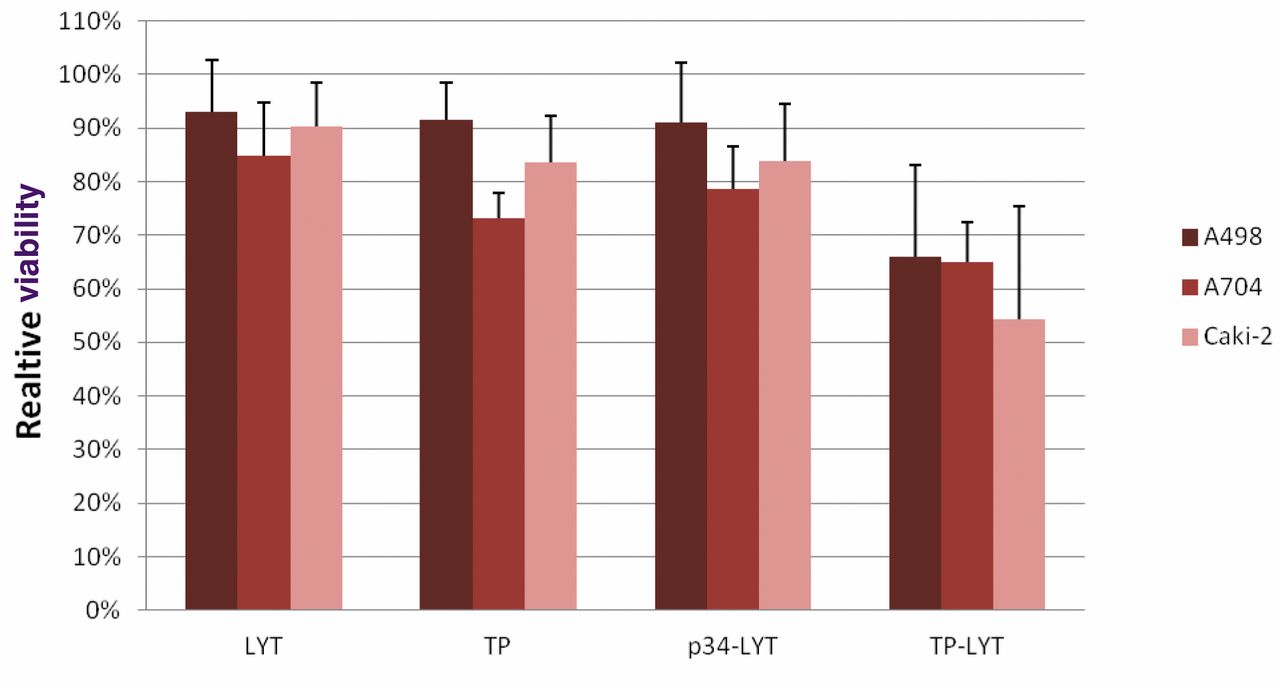
Toxicity of the bi-functional peptide towards renal cancer cells. Firstly, we designed a peptide which consists of 23 amino acids and two functional domains. The target domain (TP) was coupled to a glycin-glycin linker to the lytic domain (LYT) (Figure 1). We tested the cytotoxicity of TP-alone, LYT-alone, TP-LYT and an irrelevant peptide (p34-LYT) on renal cancer cell lines A704, A498 and Caki-2 using MTT assay. Figure 2 shows the viability after 24 h of incubation with the various peptides. LYT, TP and p34-LYT by themselves had minor effects on cell viability, whereas the toxicity of TP-LYT was significantly higher on all tumor cells (A498: LYT/TP-LYT p≤0.01, TP/TP-LYT p≤0.02, p34-LYT/TP-LYT p≤0.02, A704: LYT/TP-LYT p≤0.005, TP/TP-LYT p≤0.03, p34-LYT/TP-LYT p≤0.01; Caki-2: LYT/TP-LYT p≤0.02, TP/TP-LYT p≤0.04, p34-LYT/TP-LYT p≤0.04).
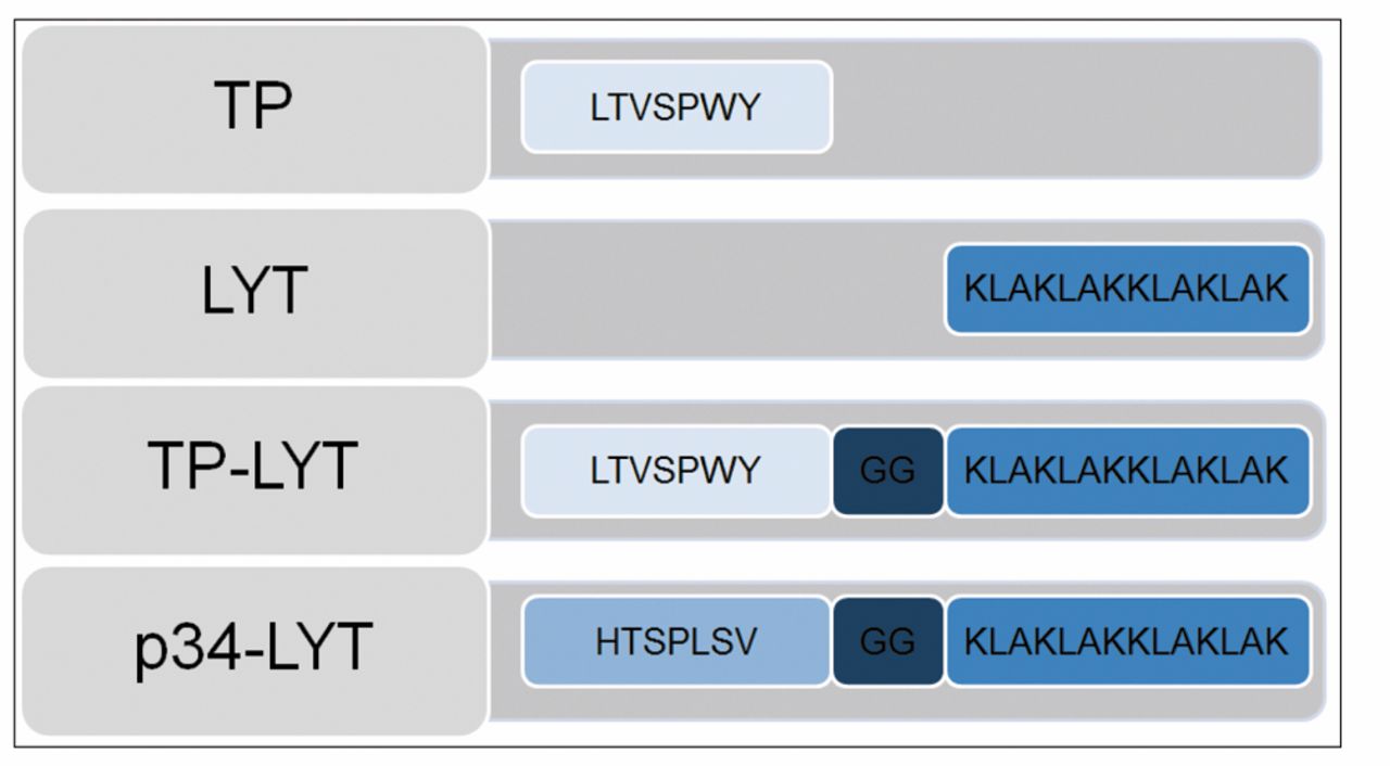
Amino acid sequence of the peptides used in this study.
Combination of WNT inhibitor and TP-LYT. Next we investigated whether TP-LYT in combination with WNT inhibitors would have an additional cytotoxic effect while remaining selective for cancer cells.
PO and TP-LYT: Figure 3A shows the effects of PO-alone, TP-LYT-alone and their combination on A704, A498, Caki-2 and CCD18Co cells. All had toxic effects on the renal cancer cell lines. Moreover, the combination of TP-LYT and PO reduced viability significantly more, compared to PO-alone and TP-LYT-alone. At the same time, it had hardly any effect on the CCD18Co cells (A704: PO/TP-LYT+PO p≤0.03, TP-LYT/TP-LYT+PO p≤0.001; Caki-2: PO/TP-LYT+PO p≤0.006, TP-LYT/TP-LYT+PO p≤0.03; A498: PO/TP-LYT+PO p≤0.0005, TP-LYT/TP-LYT+PO p≤0.003).
CIC and TP-LYT: As illustrated in Figure 3B, CIC-alone, TP-LYT-alone and the combination of both, present similar results as for the combination with PO. Each had a toxic effect by itself, but a significantly higher cytotoxic effect when combined (p≤0.05) while the CCD18Co cells remained mainly unaffected (A704: CIC/TP-LYT+CIC p≤0.006, TP-LYT/TP-LYT+CIC p≤0.00006; Caki-2: CIC/TP-LYT+CIC p≤0.0004, TP-LYT/TP-LYT+CIC p≤0.02; A498: CIC/TP-LYT+CIC p≤0.01, TP-LYT/TP-LYT+CIC p≤0.005).
EA and TP-LYT: EA-alone and TP-LYT-alone reduced cell viability; the combination of both again had a significantly higher toxicity (p≤0.05), but with this concentration the CCD18Co cells were affected in the same way as the cancer cells (Figure 3C) (A704: EA/TP-LYT+EA p≤0.03, TP-LYT/TP-LYT+EA p≤0,008; Caki-2: EA/TP-LYT+EA p≤0.0009, TP-LYT/TP-LYT+EA p≤0.02; A498: EA/TP-LYT+EA p≤0.01, TP-LYT/TP-LYT+EA p≤0.03).
Discussion
The bi-functional peptide TP-LYT is significantly more cytotoxic to renal cancer cells than the single compounds TP or LYT (Figure 2). This indicates that LYT is relatively harmless at concentrations we used when applied outside of the cell. Once internalized with the help of the target domain TP, LYT is able to induce cell death by disrupting the mitochondrial membrane. This is due to the fact that LYT has a positively-charged structure and is therefore more attracted to negatively-charged membranes, such as eukaryotic mitochondrial membranes and prokaryotic membranes, than to neutral eukaryotic plasma membranes (13, 14). Tumor cells express higher levels of anionic phospholipids, as compared to healthy cells (15, 16). This results, overall, in a more negative plasma membrane potential. While our concentrations of LYT seemed to have no effect on plasma membranes of cancer nor of healthy cells, Jäkel et al. (17) showed that LYT used at higher concentrations interacts with the plasma membrane of cancer cells and kills them effectively. In our experiments, we coupled LYT with a target domain in order to deliver LYT into the cell. Doses of TP-LYT as low as 15 μM reduced viability by 41% in A498 and 43% in Caki-2 cells. A704 cells were less sensitive to TP-LYT, as 100 μM only reduced cell viability by 23%. This raises the question whether A704 cells have a different surface structure which inhibits interaction with the target domain TP. Further research is needed to identify the receptor or surface structure by which TP binds to cancer cells. At this point we only know for sure that this interaction is independent of the HER2 status of the cell (12). We combined TP-LYT with WNT inhibitors as aberrant activation of the WNT pathway influences the initiation and progression of renal cancer (5, 6). EA and CIC have been identified as WNT/β-catenin inhibitors (23). CIC is used as topical treatment for yeast infections in humans. It inhibits metal- dependent enzymes in the cell as it is a chelator of polyvalent metal cations (e.g. Fe3+, Al3+). In addition, it was found to block the cell cycle near the G1/S phase boundary (18). PO has a similar chemical structure to CIC and is an ethanolamine salt of the hydroxamic acid derivative piroctone. It is used in cosmetic products such as anti-dandruff shampoo and is known to have bactericidal and fungicidal effects. PO penetrates the cell membrane and forms complexes with iron(III)ions, resulting in the inhibition of energy metabolism in mitochondria (9). Furthermore, it is an active collagenase inhibitor (19). The chemical structure taken together with our results suggests that PO, like EA and CIC, is a WNT inhibitor. EA is a loop diuretic agent which was once commonly used. EA has been shown to be cytotoxic towards cancer cells and to increase the cytotoxicity of other anticancer drugs (7, 20, 21). EA inhibits glutathione-S-transferase (GST) which leads to higher glutathione (GSH) levels in the cell. Increased oxidative stress was thought to be the reason for induction of apoptosis by EA. However, a study by Aizawa et al. showed that EA-induced apoptosis is independent of the GSH level (22). The antioxidant N-acetyl-L-cysteine (NAC) inhibited EA-induced cell death, while GSH levels increased in the same way. The cytotoxic mechanism of EA seems to be its direct interaction with lymphoid enhancer-binding factor-1 (LEF-1), thus resulting in the destabilization of the LEF-1-β-catenin complex. In this way, it inhibits the WNT pathway (23). The cytotoxicity of EA towards the control cells was surprising, as EA is a clinically used diuretic agent. The doses we used might have been too high, but for clarification, further experiments are needed. In summary, our results demonstrate that the combination of TP-LYT with PO, EA or CIC has synergetic effects leading to higher cytotoxicity towards cancer cells. Combining a WNT inhibitor with targeted peptides might lead to new treatment options for patients with cancer. This might not only lead to a better prognosis, but also to reduced toxic side-effects, as lower doses are consequently needed. Further research involving in vivo experiments might be of great value.

Cytotoxicity of the toxic peptide is increased by combination with the target peptide. Toxicity of the lytic peptide (LYT) and the target peptide (TP) was tested on three different renal cancer cell lines (A498, A704, Caki-2) and compared to the combination of the single peptides, the bi-functional peptide (TP-LYT) and an irrelevant peptide (p34-LYT). Cells were exposed for 24 h. N=5-7 experiments. Mean relative cell viability is given±standard error of the mean (SEM), two tailed Student's t-test was used for statistical analysis. A p-value <0.05 was considered significant.


Combination of wingless-related integration site (WNT) inhibitors and bi-functional peptide (TP-LYT) increases cytotoxicity towards cancer cells. By the use of 3-(4,5-Dimethylthiazol-2-yl)-2,5-diphenyltetrazolium-bromid (MTT) assay the cytotoxicity of WNT inhibitors piroctonolamine (PO) (A), ciclopirox olamine (CIC) (B) and ethacrynic acid (EA) (C) was tested in combination with the bi-functional peptide (TP-LYT) on three different renal cancer cell lines (A498, A704, Caki-2). Healthy colonic fibroblasts (CCD18Co) were used as control. Each combination of WNT inhibitor with TP-LYT was significantly more toxic than either one alone. TP-LYT with PO or CIC had hardly any effect on the control cells, whereas the control cells were strongly affected by EA-alone and in combination. Cells were exposed for 24-h. N=5-6 experiments. Mean relative cell viability is given ±standard error of the mean; two-tailed Student's t-test was used for statistical analysis. A p-value <0.05 was considered significant.
- Received April 3, 2013.
- Revision received April 26, 2013.
- Accepted April 29, 2013.
- Copyright© 2013 International Institute of Anticancer Research (Dr. John G. Delinassios), All rights reserved
References
In this issue


Jump to section
Related Articles
Cited By...
- In Vitro Apoptosis Induction by Fenofibrate in Lymphoma and Multiple Myeloma
- Griseofulvin Efficiently Induces Apoptosis in In Vitro Treatment of Lymphoma and Multiple Myeloma
- Clofibrate Demonstrates Efficacy in In Vitro Treatment of Lymphoma and Multiple Myeloma
- In Vitro Efficacy of Naftifine Against Lymphoma and Multiple Myeloma
- Matched-pair Analysis of Dendritic Cell Versus Targeted-therapy in Patients with Metastatic Renal Cell Carcinoma
- Flunarizine Exhibits In Vitro Efficacy Against Lymphoma and Multiple Myeloma Cells
- In Vitro Efficacy of Cinnarizine Against Lymphoma and Multiple Myeloma
- Targeting the Wnt/Beta-Catenin Pathway in Renal Cell Carcinoma