


Abstract
This study aimed to prospectively correlate clinical outcomes of advanced epithelial ovarian cancer (AEOC), with the results of in vitro chemosensitivity testing of taxol and carboplatin using the in vitro histoculture drug response assay (HDRA). A total of 104 patients with AEOC were treated with combination chemotherapy of taxol and carboplatin after primary cytoreductive surgery between 2007 and 2012 at the Asan Medical Center, Seoul, Korea. To compare chemosensitivity in the HDRA with clinical response, all patients were first categorized into two groups as either sensitive to both taxol and carboplatin (SS), or not sensitive to one or both drugs (R) based on HDRA results. The recurrence rate was much lower in the SS group compared to the R group; 29.2% vs 69.8%, respectively (p=0.02). The SS group had a significantly longer progression-free survival compared to the R group, 34.0 months vs 16.0 months, respectively (p=0.025). These results demonstrate that the HDRA prospectively correlates to clinical outcome from chemotherapy and that treatment regimens can be individualized based on the HDRA.
- Chemosensitivity
- HDRA
- histoculture drug response assay
- epithelial ovarian cancer
- taxol
- carboplatin
- prospective clinical trial
- correlation
- prediction
- individualized therapy
Ovarian cancer is the ninth most common cancer in women, often leading to death of the patient. The treatment of choice is cytoreductive surgery followed by a taxane and platinum-based combination regimen (1, 2). Early detection and complete remission is possible only in a small portion of patients due to lack of early symptoms (3-5). In addition, there is frequent recurrence and a high chemotherapy failure rate. Individual carcinomas, even of the same histological classification, have highly divergent responses to chemotherapeutic agents (1). Individual tumors may or may not respond to an agent commonly used for a particular tumor type. Because of severe side-effects, a patient often cannot be further treated after failure of the first-line regimen (2). Consequently, there is a need for better therapy design. The histoculture drug response assay (HDRA) solves many previously-encountered problems with in vitro testing of tumors. Most importantly, the HDRA maintains three-dimensional tumor-tissue histology in culture (6-14). The HDRA has been reported to be clinically useful in breast, gastrointestinal and head and neck cancer (15-20). Chemosensitivity determined by the HDRA was a strong predictor of survival in patients with non-small-cell lung cancer (21-23). The HDRA is also accurate in predicting the response to chemotherapy in patients with ovarian cancer (3). Therefore, in the present study, the HDRA was prospectively tested for correlation in advanced epithelial ovarian, fallopian, and primary peritoneal cancer to clinical outcome of first-line adjuvant chemotherapy with taxol and carboplatin.
Patients and Methods
Patients. One-hundred and forty-eight patients with epithelial ovarian cancer with International Federation of Gynecology and Obstetrics (FIGO) stage III to IV, underwent primary cytoreductive surgery followed by combination chemotherapy of taxol and carboplatin between January 2007 and December 2012 at the Asan Medical Center (Seoul, Korea). All patients gave informed consent for HDRA testing. The study was approved by the Institutional Review Board of the Asan Medical Center (S2012-1977-0001). Of the 148 patients, a total of 104 had finished cycles of adjuvant chemotherapy and were not treated with neoadjuvant chemotherapy and did not have a coexisting malignancy. The median age of those patients was 58 years (36-80 years). The median follow-up duration was 26.0 months (1.5-66.0 months). The FIGO stage distribution was as follows: three in stage IIIa, 1 in stage IIIb, 76 in stage IIIc, and 24 in stage IV. The histological type showed 84 patients with papillary serous type; 41 with serous type; four with clear cell carcinoma; and five with mixed mullerian malignant tumor, while one had undifferentiated type (Table I).
Cytoreductive surgery. Patients diagnosed with ovarian cancer underwent a complete staging operation and the tumors were classified according to FIGO. The surgical staging operation included total abdominal hysterectomy, bilateral salpingo-oophorectomy, omentectomy, appendectomy, and para-aortic and pelvic lymphadenectomy. Further, more aggressive resection of tumor-invaded organs, such as intestinal resection, colectomy, liver segmentectomy or splenectomy, was performed in selected cases with obvious tumor invasion. All 104 patients had undergone optimal debulking operations such that the size of residual tumor after surgery was less than 1 cm3.
Chemotherapy administration. Three to four weeks after surgery, all patients started adjuvant chemotherapy with taxol (175 mg/m2) and carboplatin (5 areas under the curve [AUC]), which was administered once every three weeks. Chemotherapy was halted if fever, thrombocytopenia, or neutropenia occurred.
The histoculture drug response assay. Tumor samples were obtained during cytoreductive surgery and aseptically washed in Hanks' balanced salt solution (HBSS; GIBCO, Gaithersburg, MD, USA) and minced into fragments approximately 0.5 mm in diameter. Necrotic and non-viable portions were excluded using 3-(4,5 dimethylthiazol-2-yl)-5-(3-carboxymethoxyphenyl)-2-(4-sulfo-phenyl)-2-tetrazolium (MTS; Sigma, St. Louis, MO, USA) staining. The viable cancer tissues were weighed on a chemical balance and placed onto collagen gels (Gelfoam®; Pharmacia & Upjohn, Kalamazoo, MI, USA) immersed in 1 ml RPMI-1640 medium (GIBCO) supplemented with 20% fetal calf serum (Biowest, Nuaillé, France) and drugs in 24-well plates. Six and four replicates were concurrently run for the control and treatment groups, respectively. After incubation for 72 h at 37°C with 5% CO2, 100 μl 0.06% collagenase type I (Sigma) in HBSS and 0.2% MTT (Sigma) in PBS containing 50 mM sodium succinate (Wako Ind., Tokyo, Japan) were added to each well. Plates were incubated for another 4 h, the medium removed, and 0.5 ml dimethyl sulfoxide (DMSO) (Sigma) added to each well to extract MTT formazan. Extracts from each well (100 μl) were transferred to a 96-well plate and the absorbance was measured at 540 nm using a microplate reader (VersaMax, Sunnyvale, CA, USA). The rate of inhibition of cancer-cell viability (IR) was calculated using the following equation: IR (%)=(1−mean absorbance of treated wells per gram of tumor/mean absorbance of control wells per gram of tumor)×100. In our study, the IR cut-off value for positive response was previously determined as ≥30% (17, 18).
Assessment of chemosensitivity with the HDRA and clinical response. To compare chemosensitivity of taxol and carboplatin in the HDRA with clinical response to these drugs, the 104 patients were divided into three groups by the HDRA results for each drug: drug resistant [IR greater than −0.5 standard deviation (SD) from the mean IR]; intermediate (IR between −0.5 SD and +0.5 SD from the mean IR); or sensitive (IR more than 0.5 SD from the mean IR). With this classification, all patients were further categorized into two groups as either sensitive to both taxol and carboplatin (SS), or resistant to one or both drug (R).
Clinicopathologic characteristics of the patients (N=104).
Between these two groups, inhibition rates for taxol and carboplatin, the decrease in the CA-125 level after the second and sixth cycle of chemotherapy, progression free survival (PFS) and overall survival (OS) were analyzed and compared. CA-125 levels were measured before surgery and after every cycle of chemotherapy. Imaging [(CT) or (PET/CT)] was done before surgery and after every three cycles of chemotherapy to examine the degree of disease control. PFS was defined as the interval between the date of debulking surgery and the date of imaging. In the patients without progression, PFS was defined as the duration from the date of surgery to the last follow-up. OS was defined as the interval between the date of debulking surgery and the date of death if the patient died of disease or the last follow-up date if the patient was still alive with or without disease.
Statistical analysis. Age, stage, surgical and chemotherapeutic treatment, CA-125 level, PFS and OS of each patient were all collected based on medical records. The χ2 test and Fisher's exact test were performed to compare the characteristics of patients and response rate. The Kaplan–Meier method was used for PFS and OS. Data analysis was performed using SPSS ver.19.0 (SPSS Inc., Chicago, IL, USA). A p-value less than 0.05 was considered to be statistically significant.

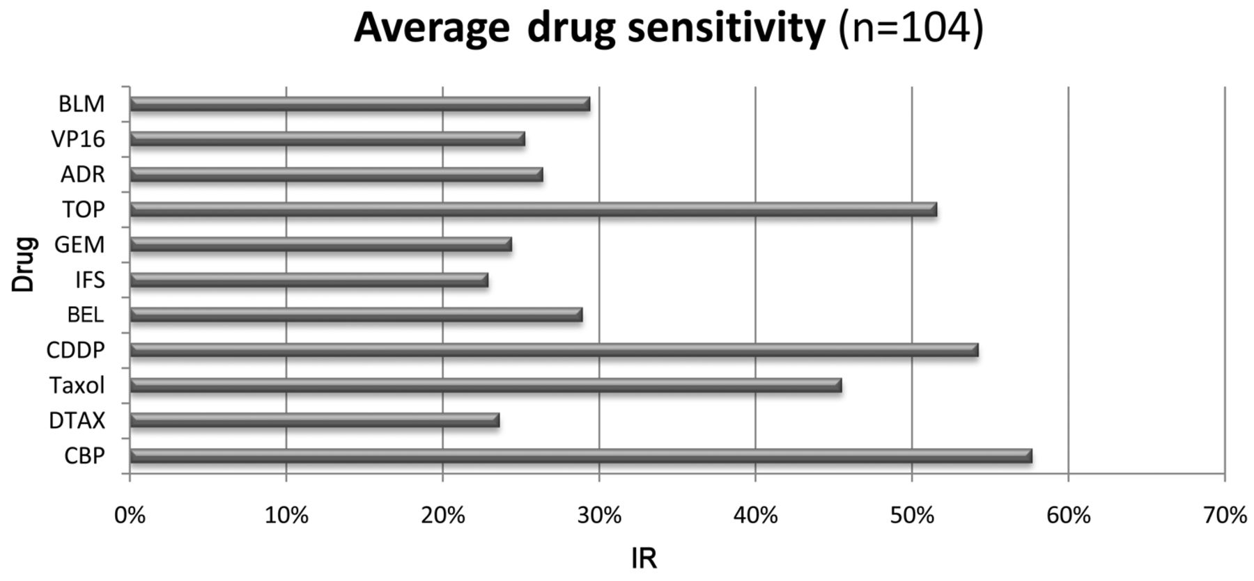
Average inhibition rate (IRs) of tumors for drugs in the HDRA for 104 ovarian cancer. BLM: Bleomycin; VP16: etoposide; ADR: adriamycin; TOP: topotecan; GEM: gemcitabine; lFS: ifosphamide; BEL: belotecan; CDDP: cisplatin; DTAX: detaxel; CBP: carboplatin.
Results
In this prospective study, 56 patients (53.8%) experienced disease recurrence and 13 patients (12.5%) died of their disease during 26.0 (1.5-66.0) months of median follow-up. The characteristics of 104 patients are shown in Table I. In vitro chemosensitivity of 11 chemotherapeutic agents was determined by the IR in the HDRA for the tumor sample obtained from each patient at surgery. The mean IRs for taxol and carboplatin, first-line for ovarian cancer, were 46% and 58%, respectively. Cisplatin, another platinum agent, and topotecan, a topoisomerase inhibitor, had IRs of 54% and 52%, respectively (Figure 1).
To compare clinical outcomes with the IRs determined in the HDRA, all patients were divided into three groups as described above. The distribution of patients was as follows: 40 patients were resistant, 24 were intermediate, and 40 were sensitive to taxol; 46 patients were resistant, 19 were intermediate, and 39 were sensitive to carboplatin. With this classification, the patients were further divided into two groups, again considering the results of both drugs: 24 patients were included in the SS group and 49 in the R group.
Table II shows the characteristics of the patients in each group. The median age (range) of the SS group was 53 (38-73) years and for the R group was 59 (39-80) years (p=0.629). The distribution of FIGO stage and the histological types were different in each group but they were not statistically significant (p=0.385 and p=0.565, respectively). The median IRs (range) for carboplatin were significantly different in each group: 82% (69-89%) in the SS group vs. 45% (4-67%) in the R group (p<0.01). The median IRs (range) for taxol were also higher in the SS group than the R group: 66% (51-90%) vs. 35% (2-50%) but not statistically significant(p=0.643).
Patients in the SS group had more tendency to have CA-125 levels below 10 U/mL after the sixth cycle of chemotherapy compared to the R group: 79.2% vs. 65.3%, respectively (p=0.23).
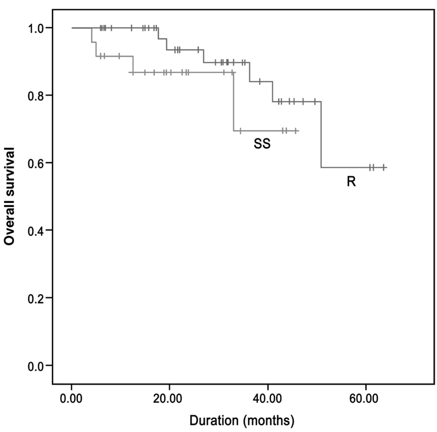
The recurrence rate was much lower in the SS group compared to the R group: 29.2% vs. 71.4%, respectively (p=0.02). The median PFS was also significantly longer in the SS group at 34.0 months compared to the R group at 16.0 months (p=0.03) (Figure 2). The median OS was not able to be evaluated due to the small number of deaths during the follow-up period as shown at Figure 3.
The median PFS appeared to be influenced significantly by carboplatin but not by taxol. Patients who were sensitive to carboplatinin in the HDRA had a longer PFS than those who were resistant at 33.0 vs. 28.0 months (p=0.01) but the median PFS did not differ whether patients were sensitive to taxol or not (35.0 vs. 32.0 months, p=0.75).
Discussion
The present clinical trial was performed prospectively in a single institution, and the results of the HDRA for both carboplatin and taxol were investigated as a predictor for clinical outcome of epithelial ovarian cancer. This study found a significant association between the in vitro HDRA chemosensitivity to carboplatin and taxol, and the PFS of patients with advanced ovarian cancer in a prospective clinical trial at a single institution. Patients whose tumors showed higher IRs for both taxol and carboplatin had a significantly longer median PFS and lower recurrence rate. This finding indicates that the HDRA can be applied clinically when choosing initial chemotherapeutic agents after cytoreductive surgery for ovarian cancer and also for second-line agents in recurrent ovarian cancer.
Comparison of clinicopathological characteristics of patients sensitive to both taxol and carboplatin (SS) (n=24), and those not sensitive to either drug (R) (n=49).
The HDRA has been shown to have a high evaluation rate of 98.8%, high specificity of 70-90%, and high clinical correlation of 80-90%, which is higher than other drug response assays and thought to be due to the maintenance of the three-dimensional tissue architecture (11, 12, 15, 20).
This study also revealed that the chemosensitivity to carboplatin but not to taxol, in the HDRA was independently associated with PFS. The two drugs have a different mechanism of action: carboplatin interacts with DNA to interfere with DNA repair, while taxol disrupts microtubules essential to mitosis.
Even though it was not statistically significant, there was a tendency for a decrease of CA-125 and the proportion of patients with CA-125 below 10 U/mL increased after the sixth cycle of chemotherapy in the sensitive group but not the resistant group. This result implies that the level of CA-125 after several cycles of chemotherapy may also be another factor for predicting the clinical outcome of advanced ovarian cancer by reflecting the degree of chemosensitivity.
Because of the short-term follow-up duration and small number of patients, this study was not able to compare the median OS between the HDRA sensitive and resistant groups. However, the study was performed prospectively in a single institution, implying that enrolled patients can be considered to receive homogeneous cytoreductive surgery and adjuvant chemotherapy.
In conclusion, results of HDRA are related to the median PFS and recurrence rate in advanced ovarian cancer, which indicate that chemotherapy can be individualized for better prognosis by selecting more effective agents, especially when the IR for first-line agents, carboplatin and taxol, are very low in the HDRA.

Kaplan–Meier survival curves showing progression-free survival of patients sensitive to both taxol and carboplatin (SS group, n=24), and patients not sensitive to either drug (R group, n=49). The median PFS was longer in the SS group than in the R group (34.0 vs. 16.0 months, p=0.03).

Kaplan–Meier survival curves showing overall survival of patients sensitive to both taxol and carboplatin (SS group, n=24), and those not sensitive to either drug (R group, n=49) (p=0.27).
- Received January 2, 2013.
- Revision received February 8, 2013.
- Accepted February 8, 2013.
- Copyright© 2013 International Institute of Anticancer Research (Dr. John G. Delinassios), All rights reserved
References
In this issue


Jump to section
Related Articles
Cited By...
- Correlation Between Clinical Outcomes and Serum CA-125 Levels After Standard Treatment for Epithelial Ovarian Cancer
- Applicability of the Histoculture Drug Response Assay to Predict Platinum Sensitivity and Prognosis in Ovarian Cancer
- Assessment of the Applicability of Integrative Tumor Response Assays in Advanced Epithelial Ovarian Cancer
- Clinical Significance of 5-Fluorouracil Chemosensitivity Testing in Patients with Colorectal Cancer
- Correlation Between 18F-FDG-uptake and In Vitro Chemosensitivity of Cisplatin in Head and Neck Cancer
- Independence of Cytotoxic Drug Sensitivity Profiles and Receptor Subtype of Invasive Ductal Breast Carcinoma Demonstrated by the Histoculture Drug Response Assay (HDRA)
- Long Noncoding RNA MRUL Promotes ABCB1 Expression in Multidrug-Resistant Gastric Cancer Cell Sublines