


Abstract
Background: Extramedullary (EM) organ impairment in patients with multiple myeloma (MM) is a rare event, occurring mostly during disease relapse after high-dose chemotherapy with autologous or allogeneic stem cell transplantation. This manifestation is commonly associated with an unfavourable outcome. Previous studies suggested a correlation between the clinical course of patients with MM and EM and the cytogenetic findings, e.g. deletion of TP53 on 17p13. Materials and Methods: We investigated patients with these rare plasma cell organ infiltrations (n=17) as well as bone lesions or soft tissue lesions, known to be a common clinical feature of MM (n=14), using a newly established method of fluorescence in situ hybridization in combination with cytoplasmic immunoglobulin staining (cIg-FISH) on paraffin-embedded sections and a specific probe for TP53 on 17p13. Results and Conclusion: The incidence of del(17)(p13) was similar in both groups but overall it was higher when compared to published data obtained from bone marrow samples and material originating from osteolyses. Further investigations on a larger patient cohort are needed in order to confirm these findings.
- Extramedullary myeloma
- cytogenetics
- TP53
- multiple myeloma
- fluorescence in situ hybridization
- cIg-FISH
- del (17)(p13)
Extramedullary (EM) organ impairment is a rare and late event in the course of multiple myeloma (MM), mostly occurring during disease relapse after high-dose chemotherapy with autologous (auto) or allogeneic (allo) stem cell transplantation (SCT). A recent report recorded an incidence of 9.3% for isolated EM relapse and a cumulative incidence of 20.4% for EM relapses among patients with MM treated with sequential auto- and allo-SCT (1). Another study with fewer patients found an even higher incidence (37%) of EM relapses after allo-SCT (2). The occurrence of EM disease is associated with a very rapid clinical progression and an unfavourable outcome due to refractoriness to chemotherapy (3). In contrast, primary extramedullary plasmacytomas (EMP) are defined as isolated and localized extraosseus plasma cell tumours without bone or bone marrow involvement and without any signs of systemic spread. They are characterized by an indolent clinical course and by radiosensitivity, with trend towards local recurrence, and only sporadic progression to MM (4). The causes for extramedullary progression or relapses in patients with MM remain unclear and there are no well-defined parameters predicting which patients are at risk of developing EM lesions. A recent study suggested a correlation between the clinical course of patients with EM organ impairment and specific genetic aberrations, especially deletion of the tumour suppressor gene TP53 on 17p13 (5). In this context, one fluorescence in situ hybridization (FISH) study of nine patients with MM, presenting with extremely rare central nervous system (CNS) involvement, demonstrated a high incidence of TP53 deletions in the bone marrow of the studied patients (eight out of nine) (6). Osteolytic myeloma lesions represent the most common clinical manifestation in patients with MM (7). Osteolyses have been investigated only in one previously published study on cytogenetic level, where deletions of TP53 were described in 7% of the studied population (8). In the current cytogenetic study, we investigated deletions of TP53 on chromosome 17p13 by applying FISH in combination with cytoplasmic immunoglobulin staining (cIg-FISH) in samples of affected organs and bone osteolyses or soft tissues of patients with MM.

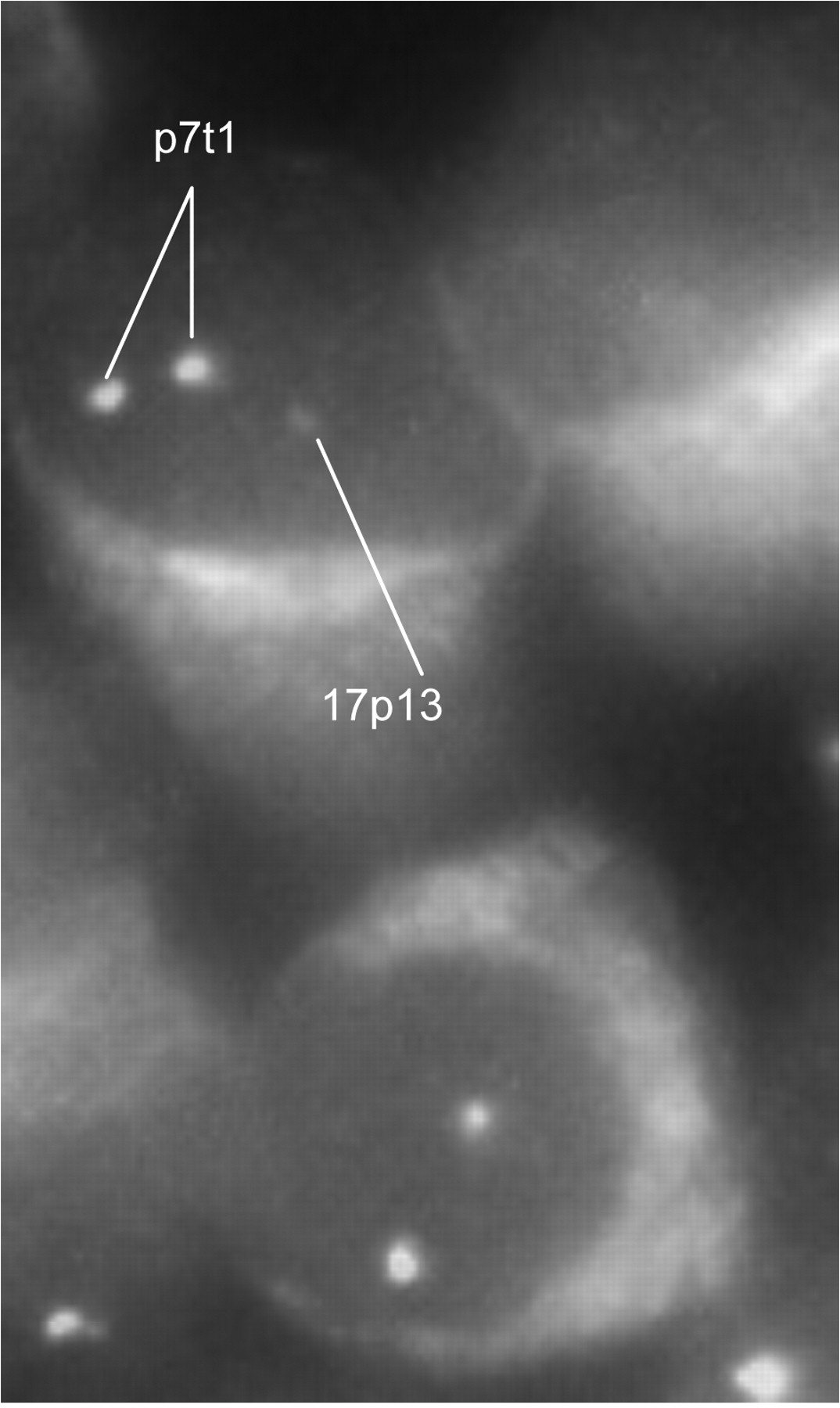
Fluorescence in situ hybridization with cytoplasmic immunoglobulin staining showing a plasma cell with normal signal counts: two signals for TP53 on 17p13 and two signals for the centromere of chromosome 7 (p7t1) in a patient with multiple myeloma with an infiltrated lymph node.
Materials and Methods
We investigated paraffin-embedded tissues of different EM organ manifestations of 17 patients with MM and of 14 patients with MM accompanied with bone or soft tissue lesions originating from a bone disease. EM organ involvements comprised of biopsies from skin, pleura, pleural effusion, uterus, liver, CNS, subcutaneous soft tissue, lymph node, and thyroid gland. The second group of material comprised of bone lesions or surrounding soft tissue, such as muscle, which was infiltrated per continuitatem. The EM manifestations in both groups were gained predominantly during disease relapse. For cytogenetic investigation of the paraffin-embedded samples, we combined FISH with a staining for the intracytoplasmatic light-chains (κ or λ) by fluorescence-labeled antibodies (cIg-FISH). We therefore advanced the well-known technique of cIg-FISH (9), successfully applied to cytospins of bone marrow aspirates, for use on paraffin-embedded samples. This gave us the opportunity to determine the fraction of plasma cells in the EM mass and to analyse them sensitively and specifically (Figure 1). For the screening of the 17p13 chromosomal region the spectrum orange probe LSI TP53 (Abbott Diagnostics, Chicago, IL, USA) was used. A specific probe for the centromeric region of chromosome 7 (p7t1) was used as a control.

Fluorescence in situ hybridization with cytoplasmic immunoglobulin staining showing deletion of TP53 in a patient with multiple myeloma with plasma cell infiltration of the liver: one signal for 17p13, two signals for p7t1 (centromere 7).
Results
Both groups showed a similar frequency of deletions of TP53. Results are summarized in Tables I and II. In the cohort of patients with soft tissue or bone involvement originating from a bone lesion, the age of presentation ranged from 45 to 80 years (mean age 66.3 years), six were female and eight were male. Three out of 14 (21%) patients had deletion of TP53 (Table I). Out of these three patients, two developed EM manifestations during disease relapse (patients 7 and 12). Unfortunately, no information regarding the disease stage of patient 9 was available. The mean age of the group of patients with MM with EM organ infiltration was 62 years (range 41 to 81 years), five were female and 10 were male. TP53 deletions were present in five out of 17 patients (29%) (Table II, Figure 2). EM manifestations occurred during disease relapse in three of the five patients (patient 2, 5 and 13) and were already present at initial diagnosis in patient 15. Information about the time of occurrence of the EM disease was not available for patient 14. Overall, we found a higher incidence of del(17)(p13) than previously reported in studies on bone marrow samples, on which it has been stated as being around 10-15% (10). Furthermore, the percentage of TP53 deletion in our MM cohort was larger than what was described in the study on material gained from osteolyses, where the investigators found an incidence of 7% (8).
Results of patients with multiple myeloma with soft tissue or bone involvement originating from a bone lesion.
Results of patients with multiple myeloma with extramedullary organ manifestation.
Discussion
EM manifestation in solid organs occurs as a late event in disease progression of MM frequently after intensive treatment modalities such as auto- or allo-SCT. It indicates an aggressive course of disease and often heralds a poor prognosis (3, 11). In contrast, osteolytic lesions represent a common clinical manifestation of myeloma at any stage of the disease (7). Information on genetic aberrations of EM plasma cell dyscrasias is sparce. One recent study investigating EMPs, as a rare subgroup, did not find any significant cytogenetic differences between EMP and MM regarding deletions of 13q14 or the translocation t(4;14) (12). Cytogenetics of EM organ manifestations or osteolyses in the context of MM have been only sporadically studied (3, 5, 6, 8). Deletion of 17p13 is known to be a late event in myeloma genesis, occurring predominantly in advanced or relapsed disease in patients with MM and is associated with a poor outcome even after allo-SCT or bone marrow transplantation (13). Therapy approaches for patients with relapsed or refractory MM include treatment with lenalidomide, bortezomib and dexamethasone (VRD), which led to an overall response rate of 68% in a recent study (14). However, the presence of TP53 deletions cannot be overcome by these novel agent-based treatments and it, thus, remains an adverse prognostic factor leading to significantly lower response rates, and shorter progression-free and overall survival (14). Some authors assume there is a correlation between the deletion of 17p13 and EM progression of MM (5, 6): A recent case report demonstrated a patient with MM with a paraspinal plasmacytoma who additionally developed an EM supraclavicular mass with pathological plasma cell infiltration during conventional chemotherapy. While a complete remission with the absence of clonal plasma cells in the bone marrow was achieved below, the EM plasmacytomas were refractory to treatment, including the high-dose chemotherapy with auto-SCT. FISH analysis showed del(17)(p13) in the EM organ impairments but not in the bone marrow (5). In this context, another FISH study of bone marrow plasma cells in nine patients with MM with CNS involvement, at the same time, reported a high incidence of TP53 deletions (89%) compared to patients without CNS involvement, where TP53 deletions ranged from 10 to 15% (6). In our study, both investigated groups had a similar incidence of deletion of TP53. However, our results revealed an overall higher occurrence of TP53 deletions compared to findings obtained from bone marrow samples and to previously published results of investigations of osteolyses (8). This latter study reported an incidence of 7% of del(17)(p13) (6 out of 88). The incidence of TP53 deletions in bone marrow samples of patients with MM in two other studies ranged from 10-16% (13, 15). Since all the patients included in our study were investigated during disease relapse, our results could be the expression of a more aggressive disease stage. Moreover, the small patient number in our groups must be taken into consideration when discussing this difference. Unfortunately, there is a lack of cytogenetic data of bone marrow cells at initial diagnosis in most of our investigated patients. Taken together, we found similar incidences of del(17)(p13) in solid organ infiltrations of EM in patients with MM and in soft tissue material infiltrated by malignant plasma cells from a bone lesion. Interestingly, we found an overall higher incidence of del(17)(p13) in comparison to published data obtained from bone marrow specimens or osteolyses themselves. These observations need to be confirmed on a larger patient cohort.
- Received February 13, 2012.
- Revision received April 10, 2012.
- Accepted April 10, 2012.
- Copyright© 2012 International Institute of Anticancer Research (Dr. John G. Delinassios), All rights reserved


