


Article Figures & Data
Figures
- Figure 1.
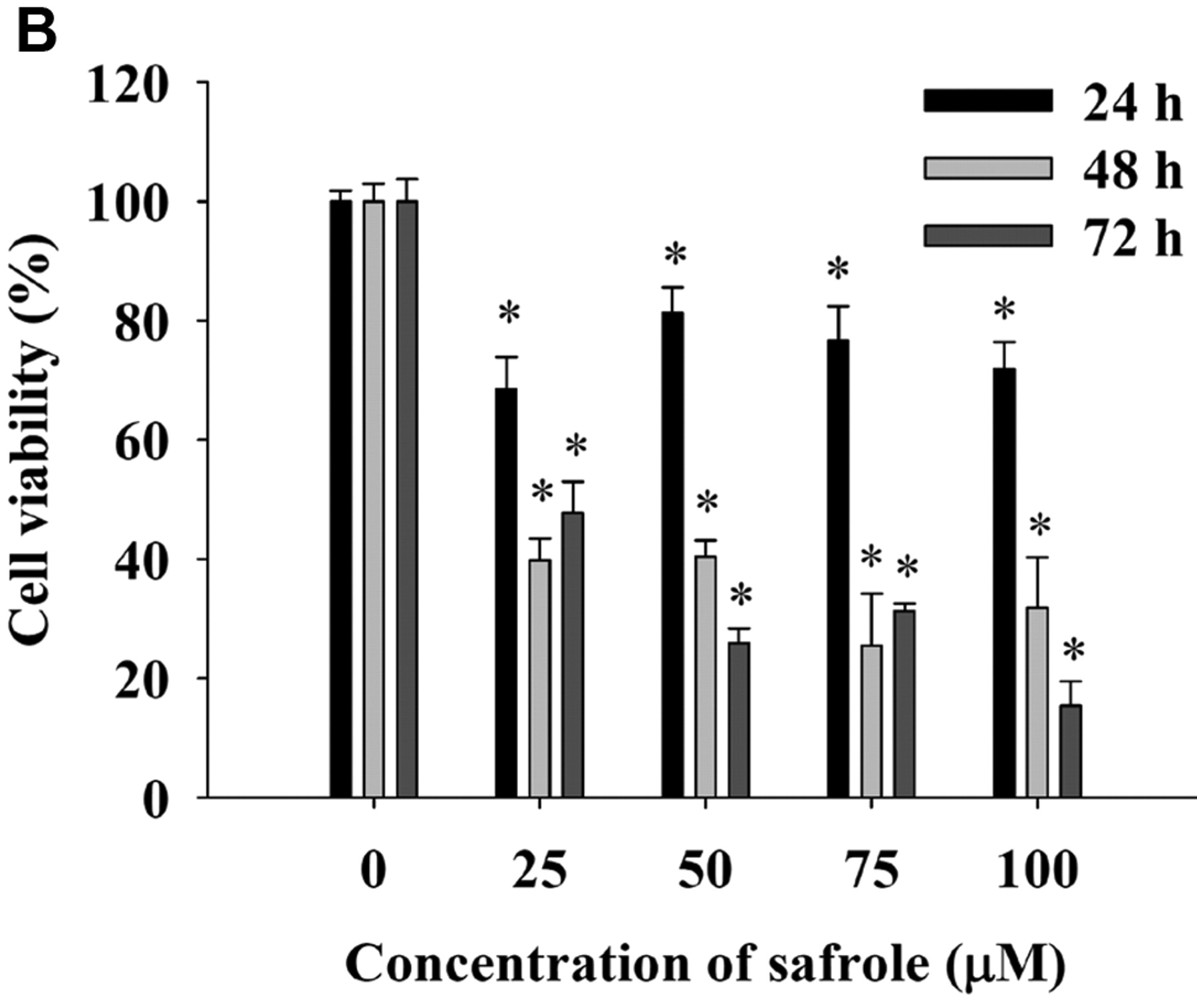
Safrole induced cell morphological changes and reduced the percentage of viable HL-60 cells. Cells were cultured in RPMI 1640 medium with 10% fetal bovine serum (FBS) with 0, 25, 50, 75 and 100 μM safrole for 24, 48 and 72 h. The cells' morphological changes were examined and photographed under phase-contrast microscopy (×200) after 24-h treatment (A). Arrows indicate cell shrinkage and rounding during cell apoptosis. The percentage of viable HL-60 cells (B) were determined as described in the Materials and Methods. Each point is the mean±S.D. of three experiments. *Significantly different at p<0.05 compared with DMSO-treated control.
- Figure 2.
Safrole affected the cell cycle distribution and the levels of the associated proteins in HL-60 cells. Cells were cultured with 0, 25, 50, 75 and 100 μM safrole for 24 h. The cells were examined and analyzed for cell cycle distribution (A) and the associated protein levels (B) by flow cytometry and western blotting as, described in the Materials and Methods. Each point is the mean±S.D. of three experiments. *Significantly different at p<0.05 compared with DMSO-treated control.
- Figure 3.
Safrole induced apoptosis and DNA damage in HL-60 cells. Cells were incubated with 0, 25, 50, 75 and 100 μM safrole for 24 h and then were harvested and were examined for apoptosis by DAPI staining (A) and for DNA damage by the comet assay (B), as described in the Materials and Methods. Results are representative of three independent experiments.
- Figure 4.
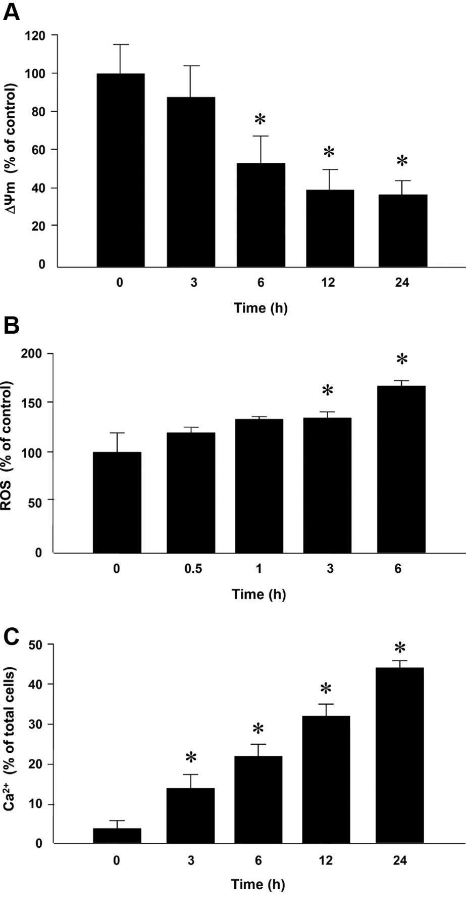
Safrole altered the mitochondria membrane potential (ΔΨm), and the levels of reactive oxygen species (ROS) and Ca2+ in HL-60 cells. Cells were treated with 75 μM safrole for indicated periods of time before being collected, and stained with DiOC6 (1 μmol/l) for ΔΨm (A), H2DCF-DA (10 μM) for ROS (B) and Indo-1/AM (3 μg/ml) for cytosolic Ca2+ (C), as described in the Materials and Methods. Each experiment was carried out with triplicate sets mean±S.D.: *Significantly different at p<0.05 compared with DMSO-treated control.
- Figure 5.
Representative western blotting showing changes in the levels of apoptosis and endoplasmic reticulum (ER) stress-associated proteins in HL-60 cells after exposure to safrole. Cells were treated with 75 μM safrole for 0, 6, 12, 24, 48 and 72 h before total proteins were prepared and determined. The levels of apoptosis-related protein expressions were estimated by western blotting analysis, as described in the Materials and Methods. BCL-2 (B-cell lymphoma 2), BAX (BCL-2-associated X protein), APAF-1 (apoptotic protease activating factor-1), cytochrome c, AIF (apoptosis-inducing factor), XIAP (X-linked inhibitor of apoptosis protein), caspase-9, FAS (fatty acid synthase), caspase-3, caspase-8, PARP (poly (ADP-ribose) polymerase), GRP78 (glucose-regulated protein 78), GADD153 (growth arrest- and DNA damage-inducible gene 153) and ATF-4 (activating transcription factor-4) and ATF6-α.
- Figure 6.
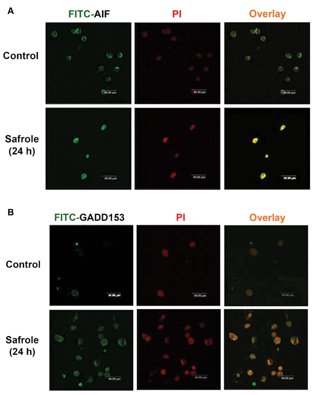
Safrole treatment resulted in translocation of apoptosis-inducing factor (AIF) and growth arrest- and DNA damage-inducible gene 153 (GADD153) in HL-60 cells. Cells were incubated with or without 75 μM safrole for 24 h, and then were fixed and stained with anti-AIF (A) and GADD153 (B) antibodies before the fluorescein isothiocyanate (FITC)-labeled secondary antibodies were used (green fluorescence) and then they were detected by a confocal laser microscope. The nuclei were stained by PI (red fluorescence). Areas of co-localization of AIF and GADD153 expression, respectively, and nuclei in the merged panels are yellow. Scale bar, 40 μm.
- Figure 7.
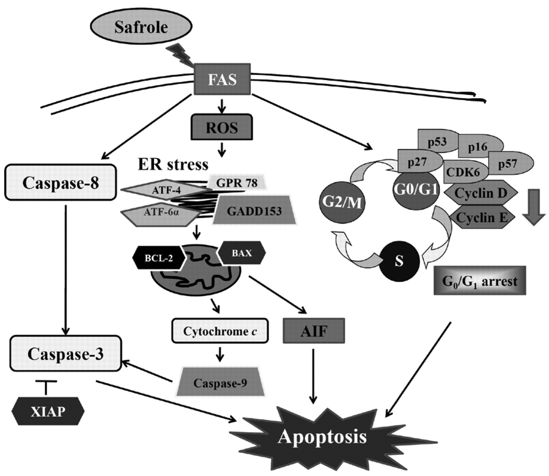
The proposed model of possible signaling pathways for human leukemia HL-60 cells after exposure to safrole.


In this issue


Jump to section
Related Articles
Cited By...
- Demethoxycurcumin-induced DNA Damage Decreases DNA Repair-associated Protein Expression Levels in NCI-H460 Human Lung Cancer Cells
- Protective Effects of Pyridoxamine Against UVC-induced Programmed Cell Death in HaCaT Cells
- Citric Acid Induces Cell-cycle Arrest and Apoptosis of Human Immortalized Keratinocyte Cell Line (HaCaT) via Caspase- and Mitochondrial-dependent Signaling Pathways
- Amentoflavone Induces Cell-cycle Arrest and Apoptosis in MCF-7 Human Breast Cancer Cells via Mitochondria-dependent Pathway