


Abstract
Background: Photochemical transformation of certain bioactive compounds for the purpose of obtaining derivatives with increased bioactivity is a prospective area of synthetic chemistry. Ecdysteroids, analogs of the insect molting hormone, which can also exert several beneficial effects in mammals including humans, contain an enone moiety in their B ring, and, as such, are good candidates for photochemical transformations. Materials and Methods: 20-hydroxyecdysone (20E), the most common ecdysteroid in Nature, and the easily obtained derivative 20-hydroxyecdysone 2,3;20,22-diacetonide (20ED), at different concentrations, were exposed to a 266 nm laser beam at an energy level of 6.5 mJ for different periods of time and evaluated for fluorescence emission during the process of irradiation. The products of irradiation were scanned from 200 to 1500 nm and then subjected to one-dimensional and two-dimensional thin layer chromatography. Results: During irradiation, progressive significant changes in the fluorescence emission spectra were noted for both compounds with time that were accompanied by changes in their UV-Vis spectra. Full conversion of both compounds was reached within 14 minutes, and both compounds yielded several major products and several minor ones representing a wide polarity range. Conclusion: The photo-transformation system described here was proven to be a useful and flexibly adjustable tool for the laser-catalyzed conversion of bioactive compounds. Due to the multi-drug resistance reversal activity of the less polar ecdysteroids, several new products are promising for being tested against various cancer cell lines. Fractionation, isolation and characterization of the irradiated products are currently in process.
For over four decades, the effect of white and ultraviolet (UV) light on the composition of complex molecules has been studied (1-4). Previously, we have shown that exposure of phenothiazines to various lasers results in major changes in their absorption and fluorescence emission spectra (5) and that with respect to the phenothiazine chlorpromazine, their antibacterial properties against a pathogenic bacterium were enhanced (5). These latter results supported the idea, previously presented, that exposure of complex compounds to lasers can rapidly alter the molecule sufficiently to yield new derivatives (6, 7) as compared to exposure to white light, which can take as long as one month (8) and for UV, weeks (9, 10).
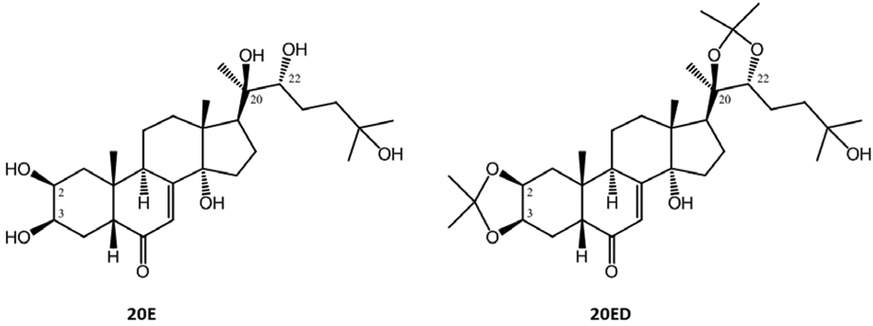
Ecdysteroids, analogs of the insect molting hormone, are frequently found in plants, where the most abundant derivative is 20-hydroxyecdysone (20E). These compounds most typically bear several hydroxyl groups on their skeleton conferring them with high polarity. On the other hand, minor ecdysteroids of much lower polarity can also be found in plants, for example 20-hydroxyecdysone 2,3;20,22-diacetonide (20ED). This compound can also very easily and economically be semi-synthesized from 20E (11). The structures of these two compounds are shown in Figure 1.
Although highly toxic in insects, in mammals (apparently lacking a well-defined ecdysteroid receptor) these compounds are of very low toxicity; the median lethal dose (LD50) for 20E in mice is 6.4 g/kg after an intraperitoneal injection (12). In fact, consumption of dietary plants containing significant amounts of ecdysteroids is held to be beneficial for human health (13). Ecdysteroids can also exert several types of bioactivities in mammals, including a mild anabolic, adaptogenic, antihyperglycemic and antihyperlipidemic activity (14, 15). Among the few reports suggesting that ecdysteroids may have specific activities on cancer cells (16, 17), of significant importance is the finding that 20ED acts on cancer cells synergistically with doxorubicin whereas its parental compound actually increases resistance to the cytotoxic agent (18).
As can be seen on Figure 1, both 20E and 20ED share a common property of ecdysteroids, which is the presence of a 7-en-6-one chromophore group on their B ring. This moiety allows these compounds to absorb UV light at a maximum of around 250 nm, and as such, makes them sensitive for photochemical transformations. UV-catalyzed transformation of enones is a special and highly promising area of modern synthetic chemistry, which permits many different types of chemical reactions to be performed, including cycloadditions with predictable stereochemistry, rearrangements via ring fragmentation, etc. (19). 20E has previously been studied for effects of UV on the generation of new species where a standard Original Hanau Photo-reactor containing an immersed Pyrex-filtered medium-pressure Hg lamp was used to yield several photo-derivatives of 20E, including an interesting dimer at acceptable yields (19% maximum) after a 4-hours exposure (20).
The concept of laser exposure of complex molecules for the purpose of generating new, potentially bioactive species is now the objective that we are pursuing. To this end, the possibility that the cancer-bioactivities of the parent compound 20E and its derivative 20ED can be modified by exposure to a high intensity laser beam is our goal and the study reported herein presents the modifications of these compounds catalyzed by a 266 nm laser beam.
Materials and Methods
Characteristics of the laser. Laser pulse repetition rate: 10 pulses per second; laser beam wavelength: λ=266 nm, which is the fourth harmonic of the Nd:YAG laser beam that emits at 1.064 μm; full-time width at half maximum of a laser pulse: 5 ns; available energy of the laser beam, variable between 5 mJ and 15 mJ; the beam diameter in the focus was set up to be the same as the inner diameter of the cuvette. During irradiation, the fluorescence emission spectrum of the exposed material was continuously monitored.
The set-up employed for exposures and monitoring of the effects of irradiation is shown in Figure 2. The laser beam is emitted by a system that applies twice second-harmonics generation (SHG) processes: on the 1064 nm beam first and 532 nm secondly. After the second SHG, the 532 nm and 266 nm were split using a diffraction grating and only the 266 nm is further used in the experiments.
The beam is optically processed so that its cross section on the sample 4 ml cuvettte covers a 9 mm2 area of the solution volume. The solution in the cuvette is agitated using a magnetic stirrer so that the reaction products that appear after interaction with the laser beam are homogeneously mixed in the solution. The beam was focused above the magnet to prevent any interference with the path of irradiation. The magnetic stirrer was set to yield approximately 700 revolutions per minute, and hence, continuous mixing was achieved without formation of eddies or bubbles.
During the exposure of the sample, laser-induced fluorescence is measured in real time in the visible spectral range using an optical fiber-spectrometer (0.65 nm resolution, Ocean Optics, Dunedin, Florida, USA) – PC chain. The absorption spectrum of the solution is previously measured using a spectrophotometer. Samples were taken from the sample cell in order to perform thin layer chromatography (TLC).
Ecdysteroids. 20E was obtained from a plant source as described previously (21); 20ED was semi-synthesized according to a published method using 5% of phosphomolybic acid as catalyst in acetone (11). Both compounds possessed a purity of over 98% by means of high-pressure liquid chromatography coupled with UV-detection (HPLC-UV). Due to the different solubility of the two compounds, the solvents used in the present experiments were methanol and chloroform for 20E and 20ED, respectively.
Implementation of the irradiation. Compounds 20E and 20ED were dissolved in their respective solvents to yield final concentrations ranging from 2 to 20 mg/l, i.e. 1-2 ml in a 4 ml cuvette. Solutions were exposed to the 266 nm laser beam for varying periods of time (minutes). During the experiments, care was taken to insure that mixing did not result in the formation of eddies or generation of bubbles. Aliquots of 10.0 μl were taken at intervals of 1, 2.5, 4.5, 8, 11 and 14 min, diluted to given concentrations identified in the text, and 10.0 μl of each interval sample was applied to a normal phase TLC plate (DC-Alufolien Kieselgel 60F254; Merck, Darmstadt, Germany) for analyzing the composition. For TLC, solvent systems of ethyl acetate-ethanol-water (16:2:1, v/v/v) (TLC1), dichloromethane-methanol (25:2) (TLC2) and toluene-acetone-ethanol-25% ammonia (100:140:32:9, v/v/v/v and 300:140:32:9, v/v/v/v) (TLC3 and TLC4, respectively) were used. Plates were evaluated under 254 nm and 366 nm UV lamps, photographed with an electronic camera, and fluorescence was also investigated with a 366 nm UV lamp after spraying the plates with concentrated H2SO4 followed by 2-3 min heating at 120°C. Start-up irradiations for up to 60 min and the analyses of products by TLC were initially carried out in order to determine minimum and maximum periods of irradiation needed to elucidate any evolutionary changes in the parental compounds per unit period of time. For a higher resolution, two dimensional (2D) TLC was utilized for testing chosen endpoints of each irradiation, where TLC1 and TLC3 were used for analyzing products of 20E in the first and second dimensions, respectively, and TLC2 and TLC4 were similarly used for products of 20ED.
At the intervals of exposure to different intensities of the 266 nm laser, the contents of the cuvette were scanned from 200 nm through 1500 nm. The scans were then compared and analyzed for any changes in the spectral properties of the contents. Unirradiated samples of each compound at the same concentrations were similarly scanned and therefore provided the basis for comparison and determination of any changes in the spectrum denoting a change in the structure of the compound and providing information on the dynamics of the conversion. Regardless of whether any changes in the optical scans were noted, the information obtained from TLC for each interval of irradiation afforded direct comparison to the scans themselves.

Structure of 20-hydroxyecdysone (20E) and 20-hydroxyecdysone 2,3;20,22-diacetonide (20ED).
The continuous irradiation of the compounds was also accompanied by simultaneous direct detection of emitted fluorescence (see Figure 2 for set-up of this component).
Results and Discussion
Analysis of aggregate formation of compounds 20E and 20ED as a consequence of concentration. Increase in the concentration of some compounds yields aggregates, which behave differently when irradiated (22). This aggregate formation is a logical consequence of approaching the upper limit of solubility. Regarding the two compounds discussed here, 20E has a solubility of around 25 mg/ml in alcohol, while 20ED can easily reach much higher concentrations in chloroform. Based on this, 20E was analyzed for its ability to form aggregates by methods previously described (22), in order to select concentrations that do not result in aggregation so that a homogenously distributed compound is presented to the laser. Due to its high molar absorbance, most of the concentrations of 20E, chosen for testing, could not be analyzed at the UV maximum wavelength, instead two off-peak wavelengths, 269.2 and 272 nm were used for analyzing aggregate formation (23, 24). Figure 3 shows the results of these tests.
As shown by Figure 3, concentrations as high as 20 mg/ml did not yield any evidence of aggregate formation. Hence, the maximum concentration of both compounds 20E and 20ED used in the study was 20 mg/L.
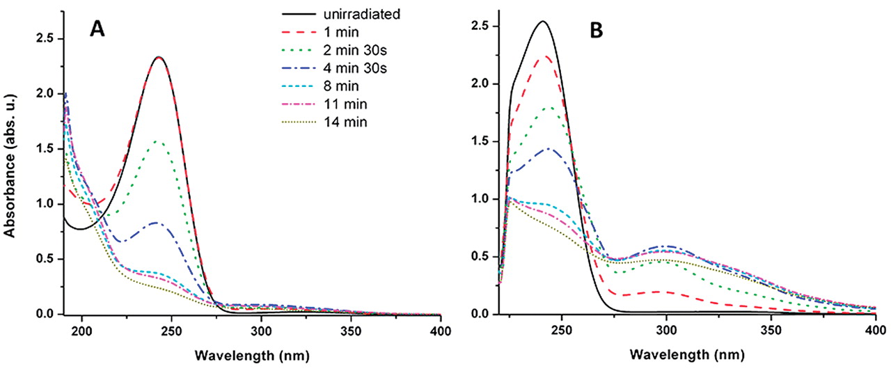
Analysis of the conversion by taking UV absorption spectra and by real-time monitoring the fluorescence emission during radiation. As our first-line analytical technique, optical scans from 200 to 450 nm served to identify any major changes produced by laser exposure of the ecdysteroids and as evident from the composite Figure 4, a change in the scans was noted as early as 1 min of exposure for 20ED and 2.5 min for 20E; evidence that the laser exposed compounds undergo further significant alterations in their structure with prolonged exposure is also apparent from Figure 4.
Fluorescent emission scans were applied as second-line monitoring of structural changes, as shown in Figure 5.
For both 20E and 20ED, the almost complete disappearance of the typical UV absorption of the parental compounds can be seen within 14 min of exposure (Figure 4). This provides strong evidence suggesting the breakdown of most of the 7-ene-6-one chromophore groups, and as noted from the gradual development of fluorescence (Figure 5), suggests the replacement of both parental compounds by another type with more extended conjugation. In the case of a longer exposure, fluorescence observed for 20ED was found to be stable for as long as 2 h post-irradiation, suggesting that most of the emission comes from photo-stable product(s), while in case of 20E, the emission of fluorescence (which was also much weaker with a more complex pattern than that of 20ED) began to decrease after 15-20 min and completely disappeared after 40 min.
Analysis of effects of irradiation on the parental compounds 20E and 20ED by TLC. The third line for analyzing the conversion of 20E and 20ED was the use of one- and two-dimensional TLC, providing valuable data on the composition of each mixture obtained at different times of irradiation. For startup, a 1 mg/ml solution of 20ED was irradiated at 6.5 mJ and ca. 5 μl aliquots were collected and analyzed by TLC at times 5, 10, 20, 30, 40 and 60 min. Results showed that the starting material experienced significant changes in its structure as early as 5 min and appeared to be completely converted at 10 min. After this, 2 mg/ml solutions of both compounds were irradiated at the same energy level for 14 min, and 10.0 μl aliquots were analyzed by TLC2. For an increased resolution, endpoints of both irradiations were also investigated by 2D-TLC. Figure 6 shows results of these analyses.

Experimental set-up. G (diffraction grating with 1302 groves/mm); optical fiber (quartz, inner core 1mm diameter); spectrometer (HR4000 Ocean Optics spectrometer with 0.65 nm resolution and 200-1100nm recording spectral range); cuvette (10 mm × 10 mm spectrophotometer cell); spectrophotometer (Perkin Elmer type 950 UV-VIS-NIR); TLC (thin layer chromatography); PC (computer for laser induced fluorescence measurements); Beam stop (optic unit to block /absorb the remaining laser beam); magnetic stirrer system to agitate the liquid sample in the cell.

Evaluation of aggregate formation for 20E at λ=269.2 nm (A) and λ=272 nm (B). The degree of polymerization is calculated as the slope of the log(c-cm) vs. log(cm) plot, where cm represents a chosen very low concentration at which the compound must exist in purely monomer form and c is the actual concentration of the sample. Aggregate formation of 20E at 20 mg/ml was found to be 1.000 as calculated from the absorption spectra at the two off-peak wavelengths, clearly showing that no aggregation occurs at this concentration. Plots for both wavelengths, as well as data of the linear regressions, are presented.

UV-VIS absorbance spectra of 20E (A) and 20ED (B) after periods of irradiation. The color code representing irradiation times for each individual spectrum is shown in A.

Fluorescence emission spectra of 20E (A) and 20ED (B) during irradiation. Color code representing the time of recording each respective spectra shown in Figure 5A is for both compounds. Note: although increase of fluorescence emission by both compounds takes place per unit period of time, emission of fluorescence by 20ED is far greater.
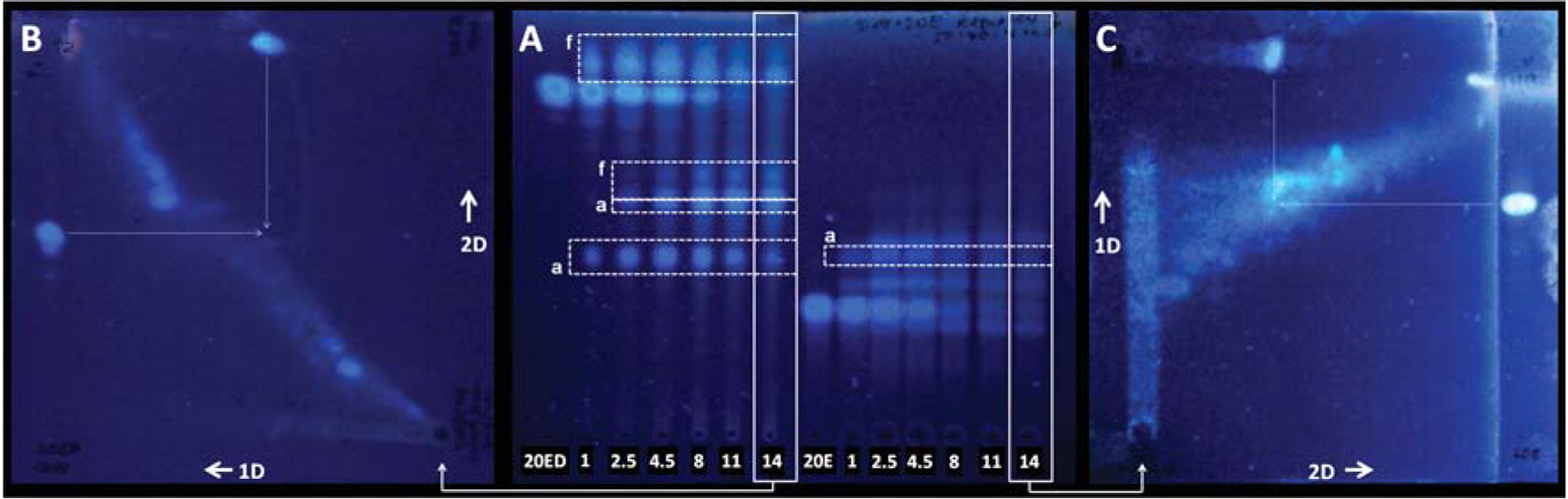
Thin-layer chromatographic analysis of the sequential products from the irradiation process of 20ED and 20E. A: Analyses of the 2 mg/ml solutions of 20ED and 20E on the left and right sides of the image, respectively, representing the process of the photolytic transformation when the compounds are exposed to a UV266 laser beam of 6.5 mJ. Numbers below the corresponding starting points represent the time in minutes when the 10 μl aliquot was collected, 20ED and 20E represent the un-irradiated compounds, a denotes products with absorbance at 254 nm and f shows those with fluorescence at 366 nm before being sprayed. B and C: 2D TLC analyses of the solutions irradiated for 14 minutes. Dashed arrows highlight the area where the spot of the remaining starting material would be expected, and 1D and 2D represent directions for the first and second dimensions of TLC, respectively.
As can be seen from Figure 6, a complete conversion for both compounds was reached in about 14 min, and the two mixtures of products exhibited a significant difference in their composition. Evaluation of the plates by three different processes (UV254, UV366 and UV366 after spraying with sulfuric acid followed by heating) also allows drawing some conclusions regarding the structural changes in the chromophore, as both parental ecdysteroids show strong absorbance at λ=254 nm, and become fluorescent at λ=366 nm only after being sprayed with a strong dehydrating reagent such as concentrated sulfuric acid or most typically vanillin-sulfuric acid. As the dashed frames show in Figure 6B, some products of 20ED seemed to present rather poor absorbance at the shorter wavelength, while weak fluorescence appeared at 366 nm. This apparent up-field shift in the UV spectrum (which is also visible from the evolution of the UV fluorescence emission spectra of 20ED during the irradiation, see Figure 5B) suggests the formation of a more extensive conjugation as compared to 20ED, which can occur for example due to the elimination of the 14-OH group. This was also visible to the naked eye: irradiation of transparent 20ED solution resulted in a mixture of yellow color. Interestingly, the presence of such products of 20E could not be observed. Considering that the two compounds are nearly identical around the chromophore group that should absorb most of the irradiation energy, the importance of the solvent cannot be ignored – the loss of a hydroxyl group seems far more favorable in chloroform than in methanol. In the case of both 20ED and 20E, the temporary appearance of a product with strong UV254 absorbance can also be seen. Although their spots appear at a nearly identical retention factor (Rf) by using TLC1, the use of other solvent systems revealed they are not the same. Regardless, the derivative of 20ED must have lost at least one of its acetonide groups to yield this compound of relatively high polarity, while it seems that most of the other products of 20ED have retained the relatively lower polarity of their parental compound as compared to that of 20E and its products.
As a next step, a scale-up for the presented photolysis was attempted. Increasing the concentration of 20ED from 2 to 20 mg/ml slightly reduced the conversion, which was overcome either by a longer irradiation time or by increasing energy. Raising the irradiation energy from 6.5 mJ to 15 mJ resulted in complete conversion of the parental 20ED in the 2 ml solution of 20 mg/ml in 14 min, while this complete conversion could also be reached by keeping the energy at 6.5 mJ but doubling the irradiation time (28 min). As for 20E, regardless of the increase of irradiation energy, the concentration of 20 mg/ml in methanol almost completely prevented conversion of the compound. The maximum concentration and volume at which this compound could still be converted was 10 mg/ml in 2 ml, at beam energy of 15 mJ. However, complete conversion required approximately 30 min. As a practical implementation of the scale-up, we performed the transformation of 400 mg of 20E and 250 mg of 20ED, in order to have raw material for the thorough chemical investigation of both product mixtures, including the isolation and structure elucidation of the individual components.
Conclusion
We have successfully set up a laser system as a powerful tool for photochemical transformation of bioactive compounds, as presented by the examples of 20E and its 2,3;20,22-diacetonide, both of which carry an α,β-enone moiety in their B ring. The described system is particularly useful for rapidly obtaining rich starting materials for bioactivity-guided isolation of novel, complex and active derivatives of similar compounds.
Due to the three simple independent analytical methods (UV-VIS spectroscopy, real-time fluorescence recording and 1D- and 2D-TLC) for the rapid screening of changes in the chemical structure of the irradiated compound(s), parameters of our system are easily and quickly adjustable for any future challenges, including the implementation of specific reactions catalyzed by UV light.
By means of our analyses, laser-catalyzed conversion of the two ecdysteroids resulted in significantly different products, each of which are potentially bioactive. Investigation of the exact chemical structures of the major and minor products of both compounds by more sophisticated spectroscopic methods (TLC-MS/MS, HPLC-MS/MS and NMR) is currently underways and bioactivity of the individual isolated compounds against cancer and efflux mediated multidrug-resistant cancer is currently ongoing and the results will soon be reported.
Acknowledgements
This work was supported by the New Hungary Development Plan (TÁMOP-4.2.1/B-09/1/KONV-2010-0005), the Baross Gábor Program (MFB-00339/2010), the ANCS (RO) project LAPLAS 3-PN 09 33 and partially by grants EU-FSE/ FEDERPOCI/SAU-MMO/59370/2004 and EU-FSE/FEDER-PTDC/BIAMIC/71280/2006 provided by the Fundação para a Ciência e a Tecnologia of Portugal. L. Amaral was supported by BCC grant SFRH/BCC/51099/2010 provided by the Fundação para a Ciência e a Tecnologia of Portugal and PTDC/SAU-FCF/102807/2008. A. Hunyadi and B. Danko were supported by STSMs within the COST Actions CM0804 and BM0701, respectively, A. Militaru and V. Nastasa were supported by projects POSDRU 107/1.5/S/80765 and POSDRU/88/1.5/S/56668, respectively.
Footnotes
- Received January 18, 2012.
- Revision received February 24, 2012.
- Accepted February 27, 2012.
- Copyright© 2012 International Institute of Anticancer Research (Dr. John G. Delinassios), All rights reserved


