


Abstract
Background: This study aimed to determine the relationship between the pre-operative metabolic tumor volume (MTV) and the disease-free survival (DFS) of patients with stage I non-small cell lung cancer (NSCLC) using F-18 2-fluoro-2-deoxy-D-glucose (FDG) positron-emission tomography–computed tomography (PET-CT) scanning. Materials and Methods: Data from sixty patients with stage I NSCLC who had undergone preoperative F-18 FDG PET-CT scanning were retrospectively analyzed. The early and late maximum standardized uptake values (eSUVm and lSUVm, respectively) were measured from attenuation-corrected F-18 FDG PET-CT images. Three MTV segmentation methods were applied as an isocontour at an early SUV of 2.5 (MTV2.5) or using fixed thresholds of either 40% (MTV40%) or 50% (MTV50%) of the maximum intratumoral F-18 FDG activity. DFS was compared by employing the Kaplan-Meier method, using the median values as cutoffs for each parameter. The log-rank test and Cox regression were performed to explore the effect of the different MTV variables on DFS. Time-dependent receiver operating characteristic (ROC) curves were created to evaluate the predictive performance. Results: During a median follow-up duration of 24 months, two patients died of disease progression, and 11 experienced recurrent tumors (eight intrathoracic tumors, two distant metastasis, and one both types of recurrences). The univariate analyses showed that pathological stage 1B, histological type of squamous cell carcinoma, male sex, maximum tumor size over 2 cm, eSUVm, lSUVm, and MTV2.5 were associated with reduced DFS. Patients who had tumors with large eSUVm or large lSUVm had a significantly lower 2-year DFS, compared with patients who had smaller tumors (65% vs. 96%, p=0.002; 63% vs. 96%, p=0.000). Patients with an MTV2.5 greater than 9.8 ml had a lower 2-year DFS than those with an MTV of 9.8 ml or greater (59 vs. 85%, p=0.02). However, multivariate analysis showed that lSUVm over 3.4 was the only parameter that exhibited an impact on DFS (p=0.05, hazard ratio=10.7), and the observed influence was marginal. Conclusion: For patients with stage I NSCLC treated with surgery, preoperative MTV parameters have a limited prognostic value for predicting DFS.
- 2-Fluoro-2-deoxy-D-glucose (F-18 FDG)
- positron-emission tomography–computed tomography (PET-CT)
- non-small cell lung cancer (NSCLC)
- metabolic tumor volume (MTV)
Stage I non-small cell lung cancer (NSCLC), including T1N0M0 and T2aN0M0 (1), is a common form of cancer. Currently, surgical resection is the first treatment option for NSCLC. Adjuvant chemotherapy is usually recommended for some patients harboring high-risk pathological features (2). Previous studies have suggested that several factors are associated with recurrence or reduced survival, such as carcinoembryonic antigen, epithelial growth factor receptor, and cytokeratin-19 fragment (3, 4). However, these factors have not been shown to be independent prognosticators (5). Recently, two investigators suggested that certain parameters measured using F-18 2-fluoro-2-deoxy-D-glucose (FDG) positron emission tomography and computed-tomography (PET-CT), such as the maximal standard uptake value (SUVmax), the metabolic tumor volume (MTV), and the total lesion glycolysis, were associated with clinical outcome of patients with advanced NSCLC (6, 7). However, which of these quantitative PET-related measurements is the best predictor of clinical outcome is unclear.
Out of these methods, the approach based on SUVmax has been the most frequently examined. Some studies indicated that a higher SUVmax of primary tumors is associated with poorer prognosis in NSCLC (7). However, the optimal threshold of SUVmax in predicting outcome remains undetermined for patients with stage I NSCLC (8). Perhaps overlapping of the SUVmax values between tumors and non-tumorous lesions might reduce its prognostic value in assessing treatment options (9). In contrast, the MTV, which can be calculated from attenuation-corrected F-18 FDG PET images as the volume of tumor with increased glycolytic activity, provides both anatomical and functional information. Several studies have suggested that the MTV may represent an independent prognostic factor in advanced NSCLC (10-13). However, investigations on the role of pre-operative MTV in early-stage NSCLC are scant. We hypothesized that the pre-treatment MTV, as measured using F-18 FDG PET-CT, is associated with survival of patients with NSCLC. Thus, we conducted our retrospective study to compare the predictive values of pre-operative SUVmax and various MTV parameters
Materials and Methods
Patient population. Between January 2009 and February 2011, 60 patients newly-diagnosed with stage I NSCLC who underwent curative surgical resection at China Medical University Hospital were enrolled in our retrospective study (Institutional Review Board certificate DMR99-IRB-010-1). They had all undergone an F-18 FDG PET-CT scan before resection surgery. The interval between the acquisition of images and the commencement of radiotherapy was less than four weeks. Each patient had an unremarkable serum glucose level before the PET-CT was performed. The pathological stage was evaluated using the tumor-node-metastasis (TNM) method (1). The cohort data are listed in Table I.
PET-CT imaging protocol. Our PET-CT procedure has been previously described (14). All patients were instructed to fast for at least 4 h before the PET-CT scan. Patients were injected intravenously with 370 MBq of F-18 FDG, and rested in a supine position in a quiet, dimly lit room. PET-CT scanning was initiated at approximately 45 min after the F-18 FDG injection using a Discovery STE instrument (GE Healthcare, Milwaukee, WI, USA). Delayed PET-CT images were obtained at approximately 70 min after the F-18 FDG injection. PET emission images were obtained from CT scans at 2 min per field of view in the 3-D acquisition modes. The initial CT images were reconstructed onto a 512×512 matrix with a section thickness of 3.75 mm, reconstructed onto a 128×128 matrix, and converted into 511-keV-equivalent attenuation factors for correction of the corresponding PET emission images. CT images were acquired twice for every patient. The SUV was defined as tracer activity in the target region per unit mass divided by the amount of injected radioactivity per unit body mass. The SUVmax of the primary tumor in both early and delayed F-18 FDG PET-CT images was obtained, and were abbreviated as eSUVm and lSUVm, respectively.
MTV definition and measurement. MTV values were measured from attenuation-corrected F-18 FDG-PET-CT images using an SUV-based automated contouring program (Advantage Workstation Volume Share version 2, GE Healthcare). Initially, the F-18 FDG-PET data were introduced into the workstation in a DICOM format, and these images were reviewed to localize the target lesions that had been confirmed by two nuclear medicine physicians. MTV was defined as the sum of the metabolic volumes of the primary tumors. To define the contouring margins around the tumor, we used an eSUVmax of 2.5 (MTV2.5), 40% of the eSUVmax (MTV40%), and 50% of the eSUVmax (MTV50%). The MTV40% and MTV50% were defined as the volume greater than a fixed threshold of 40% and 50% of the maximum intratumoral activity, respectively. The volume boundaries were automatically drawn to incorporate each target lesion in the axial, coronal, and sagittal PET-CT.
Treatment and follow-up. Following curative surgery, 16 of the 22 patients with stage IB disease were treated with adjuvant chemotherapy. The regimens used for these 16 patients were carboplatin plus gemcitabine (n=7), cisplatin plus gemcitabine (n=3), docetaxel (n=3), and oral Tegafur-Uracil (n=3). The follow-up examinations were performed every 1-2 months over the first 2 years and every 3-4 months thereafter. The definitions of local recurrence and distant recurrence were based on chest CT scanning, F-18 FDG PET-CT analysis, or pathological findings.
Statistical analysis. In this study, the median values for the MTV2.5, MTV40%, MTV50%, eSUVm, and lSUVm were used as cutoff points for dichotomization of the patient chort. Correlations between MTV and SUVmax were examined using Pearson's correlation, with the level set at 0.01. Because we sought to identify the impact of pre-treatment PET-CT parameters on cancer recurrence only, the study end-point was disease-free survival (DFS). DFS was measured from the date of diagnosis to the date of recurrence or last clinical follow-up. We analyzed MTV2.5, MTV40%, MTV50%, eSUVmax, lSUVmax, and pathological stage as categorical variables. Accordingly, the DFS curves were determined using the Kaplan–Meier method. The log-rank test and Cox regression were performed to explore the relationships between the categorical variables and DFS. The optimal predictive values of these parameters for disease recurrence were calculated by ROC curve analysis. p-Values less than 0.05 were considered statistically significant.
Results
SUVmax and MTV measurement. The mean eSUVm±SD was 3.2±2.3 (median=2.5; range=0.6-10.8), whereas the mean lSUVm±SD was 3.8±2.8 (median=3.2; range=0.6-13.4). The MTV2.5 ranged from 1.08 to 78.2 ml (median=9.8 ml, mean=11.4±12.9 ml). The mean MTV40% was 7.3±5.9 (median=5.8; range=1.7-39.0), and the mean MTV50% was 5.6±4.3 (median=4.4; range=1.2-28.0). Our data did not reveal significant correlations between the eSUVm and MTV parameters (r=0.1 for MTV2.5, p=0.46; r=0.31 for MTV40%, p=0.02; r=0.32 for MTV50%, p=0.01).
Patients' characteristics.
Treatment outcome. The median follow-up duration was 24 months (range 8-36 months). Two patients died of disease progression, and 11 experienced recurrent tumors, including eight with intrathoracic tumors, two with distant metastasis, and one with both types of recurrence. Overall, the 2-year DFS was 81%.
Prognostic value of PET-related parameters. Table II shows a summary of the effect of the tumor and the treatment-related parameters on DFS by univariate and multivariate analyses. In the univariate analysis, pT1b, squamous cell carcinoma, male sex, maximum tumor size greater than 2 cm, eSUVm, lSUVm, and MTV2.5 were associated with reduced DFS.
As shown in Figures 1A and B, patients who had tumors with large eSUVm or large lSUVm had significantly lower 2-year DFS compared with patients who had smaller tumors (65% vs. 96%, p=0.002; 63% vs. 96%, p=0.000). Figure 2 shows that patients with an MTV2.5 greater than 9.8 ml had an inferior 2-year DFS than those with smaller MTV2.5 volumes (59% vs. 85%, p=0.02). However, multivariate analysis showed that lSUVm over 3.4 was the only parameter that exhibited an impact on DFS (p=0.05, hazard ratio=10.7), and the observed influence was marginal. Overall, lSUVm yielded the best prediction for tumor recurrence. When using an lSUVm greater than 3.4 to predict disease recurrence, the sensitivity, specificity, positive predictive value, the negative predictive value, and the accuracy were 100%, 63.2%, 37.9%, 100%, and 70%, respectively.
Result of univariate, multivariate analysis for disease-free survival.
To minimize the limitation of the MTV definition in some tumors with a lower eSUVmax, an additional ROC analysis was performed for 41 of the participants who had an eSUVm greater than 2.0. The results in this subgroup also indicated that the MTV methods did not exhibit a higher predictive value than the SUVmax approach.
Discussion
Beyond the use of TNM staging, there is a need for additional prognostic markers to identify patients who are more appropriate for adjuvant therapy of early-stage NSCLC. Currently, SUVmax and MTV have been examined as prognosticators across the PET-related parameters (10-14). This study is a preliminary investigation comparing the predictive value of each in early and delayed SUVmax, as well as various MTV parameters. Theoretically, SUVmax represents the highest activity of a single pixel within the tumor, whereas the MTV represents the area where the activity of the voxels is above a selected value. The application of MTV seems more promising because MTV may combine information on tumor burden and cancer aggressiveness. Although some investigators have suggested that MTV is a useful prognostic for survival in NSCLC patients (10-14), our study failed to identify an independent predictor for DFS in patients with operable stage I NSCLC.
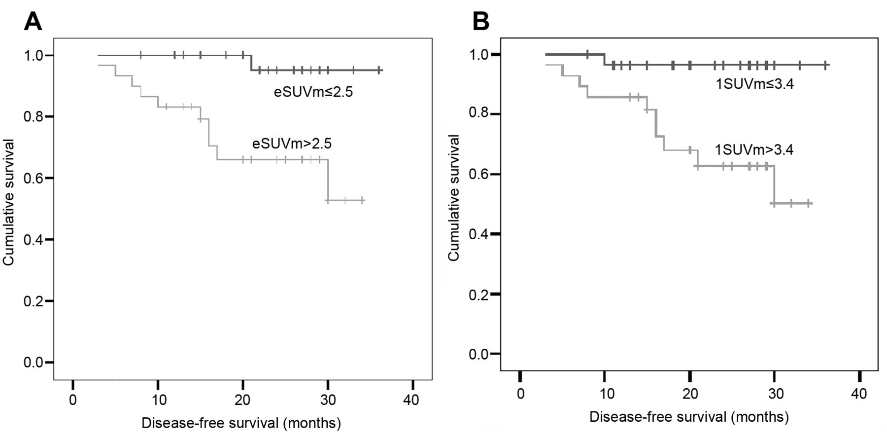
A: Disease-free survival curves according to early standardized uptake values (eSUVm). Data represent eSUVm >2.5 and eSUVm ≤2.5. B: Disease-free survival curves according to delayed SUVmax (lSUVm). Data represent lSUVm >3.4 and lSUVm ≤3.4.
A recent study (14) that enrolled patients with stage I to IIIA NSCLC, undergoing surgery indicated that MTV2.5 was a significant prognostic factor for DFS, and 42% of patients were categorized as stage II to IIIA disease. Consequently, the measured values of SUVmax or MTV would have been greater than ours. Because small tumor volume is usually associated with relatively smaller MTV, the predictive effect of MTV may be diminished because of the limitation of spatial resolution. Nonetheless, an interesting question emerges as to which threshold values for real tumor volume or SUVmax may increase the sensitivity of the predictive power of MTV using current PET-CT technologies. Future work should focus on the determination of an optimal threshold value.
There were several limitations to our study. Firstly, the median follow-up duration may not have been long enough, and further subsequent recurrences may not have been identified, resulting in the lack of affirmative findings based on the multivariate analysis. Secondly, some patients had a low SUVmax value or a small tumor volume. Thus, some technical constraints existed after measuring the MTV parameters. Regardless, the subgroup analysis did not show a contradictory result after excluding those with eSUVm less than 2.0. Nevertheless, our study represents a clinical circumstance in which the MTV approaches were restrained.

Disease-free survival curves based on metabolic tumor volume2.5 >9.8 ml and MTV2.5 ≤9.8 ml.
Conclusion
For patients with stage I NSCLC treated with surgery, pre-operative MTV parameters have a limited prognostic value for DFS. Further studies are needed to clarify the threshold values for tumor volume or SUVmax to increase the sensitivity of the predictive power of MTV measurements.
Acknowledgements
The study was partly supported by the study projects DMR-101-062 and DMR-101-080 of the China Medical University Hospital; Taiwan Department of Health Clinical Trial and Research Center and for Excellence (DOH101-TD-B-111-004), Taiwan Department of Health Cancer Research Center for Excellence (DOH101-TD-C-111-005); and International Research-Intensive Centers of Excellence in Taiwan (I-RiCE) (NSC101-2911-I-002-303).
Footnotes
-
↵* These Authors contributed equally to this work.
-
Conflicts of Interest
All Authors declare they encountered no actual or potential conflicts of interest in conducting this study.
- Received August 3, 2012.
- Revision received October 8, 2012.
- Accepted October 9, 2012.
- Copyright© 2012 International Institute of Anticancer Research (Dr. John G. Delinassios), All rights reserved


