


Abstract
Despite frequent use of topoisomerase inhibitors (TIs) as antitumor agents, their application to oral squamous cell carcinoma (OSCC) has not been reported. We investigated three inhibitors of topoisomerase I [camptothecin, irinotecan, SN-38 (active metabolite of irinotecan)] and two inhibitors of topoisomerase II (etoposide, teniposide) for their cytotoxicity towards a total of 15 human tumor cell lines and normal cultured cells. All TIs exhibited higher cytotoxicity towards tumor cell lines (OSCC, glioblastoma, myelogenous leukemia) as compared with normal mesenchymal (gingival fibroblast, pulp cell, periodontal ligament fibroblast) and epithelial cells (skin keratinocytes). Among TIs, SN-38 had the highest cytotoxicity towards OSCC cell lines, with a tumor specificity index of 1321 compared to mesenchymal cells and 22 compared with epithelial cells. SN-38 induced different types of cell death in two OSCC cell lines: apoptosis (caspase-3 activation and internucleosomal DNA fragmentation) in HSC-2 cells and autophagy (formation of autophagosome and secondary lysosome) in HSC-4 cells. The cell death of HSC-2 and HSC-4 cells was significantly inhibited by pre-treatment with caspase inhibitor (Z-VAD-FMK) and autophagy inhibitors (3-methyladenine, bafilomycin A1), respectively. The present study demonstrated that SN-38 is highly cytotoxic to OSCC cell lines, regardless of the type of induced cell death, suggesting its future application for chemotherapy of OSCC.
- SN-38
- irinotecan
- oral sqamous cell carcinoma
- apoptosis
- autophagy
- topoisomerase inhibitors
- HSC-2
- HSC-4 cells
DNA topoisomerases are nuclear enzymes that reduce torsional stress in supercoiled DNA, allowing for selected regions of DNA to become sufficiently relaxed to permit its replication, recombination, repair, and transcription (1). Two classes of topoisomerases (TOPOI and TOPOII) are known to mediate DNA strand breakage and resealing, and both have become targets of cancer chemotherapy (2-5). TOPOI have been reported to induce apoptosis (characterized by caspase activation and DNA fragmentation) in hepatocellular carcinoma Huh7 cells (6, 7), whereas TOPOII inhibitors (etoposide, teniposide) induced apoptosis in colorectal cancer cells (8, 9). However, to our knowledge, no report has been published about the clinical application of topoisomerase inhibitors in patients with oral squamous cell carcinoma (OSCC), possibly due to the lack of in vitro data of their antitumor potential against OSCC cell lines.
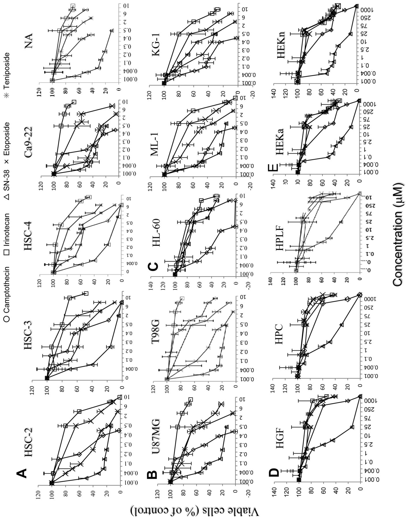
In order to test the possibility of future application of topoisomerase inhibitors for OSCC patients, in the present study we investigated the cytotoxicity of TOPOI inhibitors (camptothecin, irinotecan and SN-38, an active metabolite of irinotecan) and TOPOII inhibitors (etoposide, teniposide) (Figure 1) towards ten human tumor cell lines (OSCC: HSC-2, HSC-3, HSC-4, Ca9-22 and NA; glioblastoma: T98G and U87MG; myelogenous leukemia: HL-60, ML-1 and KG-1) and five human normal cells [oral mesenchymal: gingival fibroblast (HGF), pulp cell (HPC), periodontal ligament fibroblast (HPLF); and epithelial skin keratinocytes: HEKa and HEKn].
There are at least three types of cell death, apoptosis: type I programmed cell death characterized by blebbing, chromatin condensation, internucleosomal DNA fragmentation and the loss of cell surface microvilli; autophagy: type II programmed cell death characterized by the formation of autophagosomes and autophagolysosomes engulfing the broken organelles; and necrosis: characterized by swelling of cells and organelles (10, 11). Since the present study demonstrated that among five topoisomerase inhibitors, SN-38 exhibited the highest cytotoxicity towards OSCC cell lines, we also investigated the type of cell death induced by SN-38 in two OSCC cell lines (HSC-2 and HSC-4).

Chemical structure of topoisomerase I and II inhibitors.
Materials and Methods
Materials. The following chemicals and reagents were obtained from the indicated companies: Dulbecco's modified Eagle's medium (DMEM) from GIBCO BRL, Grand Island, NY, USA; fetal bovine serum (FBS) from JRH Bioscience, Lenexa, KS, USA; RPMI-1640, 3-(4,5-dimethylthiazol-2-yl)-2,5-diphenyltetrazolium bromide (MTT), camptothecin, SN-38, etoposide, teniposide, Hank's balanced salt solution (HBSS) and 3-methyladenine (3-MA) from Sigma Chemical Co., St. Louis, MO, USA); irinotecan from Toronto Research Chemicals Inc., Toronto, Canada; topotecan from LKT Lab. Inc. USA; pan-caspase inhibitor (Z-VAD-FMK) from Biomol, Enzo Life Science, Plymouth Meeting, PA, USA; bafilomycin A1 and dimethyl sulfoxide (DMSO) from Wako Pure Chemical Industries Ltd. Osaka, Japan.
Cell culture. HL-60 cells were provided by Professor K. Nakaya, HSC-2, HSC-3, HSC-4, NA and Ca9-22 cells were provided by Professor M. Nagumo and T98G and U87MG cells were provided by Dr. M. Iida, Showa University, Japan; ML-1 and KG-1 cells were provided by Professor K. Takeda, Tokyo University of Science. Normal oral cells (HGF, HPC and HPLF) were prepared from the periodontal tissues, according to the guidelines of the Intramural Board of Ethics Committee (no. A0808). Since normal oral cells have a limited lifespan of about 40 population doubling levels (PDL) (12), they were used at 10-14 PDL. Human skin keratinocytes (HEKa, HEKn) were purchased from KURABO Ind. Ltd., Osaka, Japan and cultured in HuMedia-KG2 supplemented with insulin, human recombinant epidermal growth factor (EGF), hydrocortisone, gentamicin, amphotericin B and bovine pituitary extract (BPE), as instructed by the supplier. HL-60, ML-1 and KG-1 cells were cultured in RPMI 1640 medium supplemented with 10% heat-inactivated FBS, penicillin G (100 units/mi) and streptomycin sulfate (100 μg/ml) in a humidified atmosphere with 5% CO2. All other cells were cultured in DMEM supplemented with 10% heat-inactivated FBS and antibiotics.
Assay for cytotoxic activity. Near-confluent cells were treated for the indicated times with or without (control) different concentrations of each inhibitor. The relative viable cell number of adherent cells was then determined by the MTT method. In brief, the cells were washed once with PBS(−), and incubated for 4 h with 0.2 mg/ml of MTT in the culture medium. After removing the medium, the reaction product, formazan, was dissolved in 0.1 ml of DMSO, and the absorbance (the relative viable cell number) was measured at 540 nm by a microplate reader (Multiskan Bichromatic Labsystems, Helsinki, Finland). The viability of suspended cells, namely, HL-60, ML-1 and KG-1, was determined by cell counting with a hemocytometer, after staining with 0.15% trypan blue in culture medium. The 50% cytotoxic concentration (CC50) was determined from the dose–response curve and the mean CC50 for each cell type was calculated from 3-6 independent experiments. The tumor specificity index (TS) (13) was calculated according to the following equations:


Assay for DNA fragmentation. HSC-2 cells (6×10⁴) were inoculated on a 6-well plate (Falcon) and incubated for 48 h to allow for cell attachment. Cells were treated for 6 or 24 h with 0, 2, 4 or 8 nM SN-38 in fresh culture medium. After washing once with phosphate-buffered saline without calcium and magnesium ion [PBS(−)], cells were collected by scraping with a rubber policeman on ice, and pelleted by centrifugation (1,000 ×g, 3 min). They were then lysed with 50 μl lysis buffer [50 mM Tris-HCl, pH 7.8, 10 mM EDTA-2Na, 0.5% (w/v) sodium N-lauroylsarcosinate] and incubated for 2 h at 50°C with 0.4 mg/ml RNase A and 0.8 mg/ml proteinase K. DNA was extracted with 50 μl NaI solution (40 mM Tris-HCl, pH 8.0, 7.6 M NaI, 20 mM EDTA-2Na) and then 250 μl of ethanol were added. After centrifugation for 20 min at 20,000 ×g, the precipitate was washed with 1 ml of 70% ethanol. DNA was dissolved in TE buffer (10 mM Tris-HCl, pH 7.5, 1 mM EDTA) and applied to 2% agarose gel electrophoresis in TBE buffer (89 mM Tris-HCl, pH 8.0, 89 mM boric acid, 2 mM EDTA). A DNA molecular marker (Bayou Biolabs, Harahan, CA, USA) and DNA from HL-60 cells induced to apoptosis by UV irradiation (6 J/m2/min, 1 min), followed by 3 h of incubation in regular culture medium, were run in parallel as a positive control (14). After staining with ethidium bromide, DNA was visualized by UV irradiation, and photographed by a CCD camera (Bio Doc-It, UVP, Inc., Upland, CA, USA).
Assay for caspase activation. HSC-2 and HSC-4 cells (6×10⁵) were inoculated onto 100-mm dishes (Falcon) and incubated for 24 h to allow for cell attachment. HSC-2 cells were treated for 6 or 24 h with 0, 2, 4 or 8 nM SN-38 in fresh culture medium. HL-60 cells were incubated for 6 h without or with 1 μg/ml actinomycin D as a positive control of apoptotic cells. These cells were washed twice with PBS(−) and lysed in lysis solution [50 mM Tris-HCl (pH 7.5), 0.3% NP-40, 1 mM dithiothreitol (DTT)]. After being placed for 10 min on ice and centrifugation for 20 min at 15,000 ×g, the supernatant was collected. Protein in the lysate was determined by the Protein Assay Kit (Bio-Rad Laboratories, Hercules, CA, USA). The lysate (50 μl, equivalent to 150 μg protein) was incubated with 50 μl lysis solution containing substrates for caspase-3 (DEVD-p-nitroanilide [pNA], caspase-8 (IETD-pNA) or caspase-9 (LEHD-pNA) for 4 h at 3°C. The absorbance of the liberated chromophore pNA was measured at 405 nm using a microplate reader (15).
Terminal deoxylnucleotidyl transferase dUTP nick- end labeling (TUNEL) assay. HSC-2 and HSC-4 cells (5×10⁴/well) were inoculated on a 8-well chamber slide (Nalge Nunc International, N. Aurora, IL, USA) and incubated for 48 h. Cells were treated for 6 or 24 h with SN-38 (8 nM) in fresh culture medium, and then assayed for the DNA cleavage by the TUNEL method using an in situ apoptosis detection kit (Takara Bio INC. Ltd., Shiga, Japan). The treated cells were washed three times with PBS(−), fixed with 100 μl freshly-prepared PBS(−) containing 4% formaldehyde, and incubated at room temperature for 30 min. The cells were then washed three times with 200 μl PBS(−), and permeabilized with 100 μl of PBS(−) containing 0.1% sodium citrate and 0.1% Triton X-100®, and left for 2 min on ice. Cells were then washed twice with PBS(−). The cells were incubated for 1 h at 37°C with 50 μl of TUNEL reaction mixture (45 μl labeling solution and 5 μl enzyme solution) in a humidified dark chamber and observed under laser scanning microscopy (16).
Observation of fine cell structure. HSC-2 cells were treated for 0, 12, or 24 h with 8 nM SN-38. Cells were then washed once with PBS(−), and fixed with 2% glutaraldehyde. The cells were post-fixed in 1% osmium tetraoxide-0.1 M cacodylate buffer (pH 7.4) at 4°C, dehydrated, and embedded in Araldite 502 (CIBA-GEIGY, Basel, Switzerland). Fine sections were stained with uranyl acetate and lead citrate, prior to being analyzed under a JEM-1210 transmission electron microscope (JEOL, Tokyo, Japan) at an accelerating voltage of 100 KV (13).
Statistical analysis. Experimental values are expressed as the mean±standard deviation (SD). Statistical analysis was performed by using the Student's t-test. A p-value <0.05 was considered to be significant.
Results
Tumor-specificity of topoisomerase inhibitors. Both TOPOI inhibitors (camptothecin, irinotecan, SN-38) and TOPOII inhibitors (etoposide, teniposide) dose-dependently reduced the viable cell number of OSCC, glioblastoma, myelogenous leukemia, normal oral mesenchymal and epithelial skin keratinocytes (Figure 2). However, their cytotoxicity towards tumor cell lines (Figure 2A-C) was much higher than the one towards normal mesenchymal and epithelial cells (Figure 2D and E, respectively). Among the inhibitors, SN-38 exhibited the highest cytotoxicity towards OSCC cell lines (CC50=0.0016-0.0039 μM), with a TS value of 1321 compared to mesenchymal cells, and 22 compared with epithelial cells (Table I).
Although camptothecin and teniposide were two orders of magnitude less cytotoxic towards OCSS cell lines (CC50=0.18-0.59 μM), as compared with SN-38, they had higher TS values of 2961 and 3190 compared to mesenchymal cells, and 372 and 321 compared with epithelial cells, respectively (Table I). Irinotecan had the least cytotoxicity towards OSCC cell lines (CC50=88 μM), with a TS value of 23 compared to mesenchymal cells and 3 compared with epithelial cells (Table I), possibly due to the lack of esterase activity in OSCC cell lines.
Type of cell death induced by SN-38. SN-38 induced caspase-3 activation in HSC-2 cells, but not in HSC-4 cells (Figure 3A). Similarly, SN38 induced internucleosomal DNA fragmentation in HSC-2 cells, but not in HSC-4 cells (Figure 3B). SN-38 induced the production of TUNEL-positive cells (another apoptosis marker) in HSC-2 cells, but not so clearly in HSC-4 cells (Figure 4).
Cytotoxic activity of topoisomerase I and II inhibitors towards human normal and tumor cells. Each value represents the mean±S.D. from three independent experiments.
Electron microscopy of HSC-2 cells treated with SN-38 is shown in Figure 5. In HSC-2 cells cultured for 12, or 24 h with 8 nM SN-38, vacuolated mitochondria containing electron-lucent matrix were observed (Figure 5A). The morphology of nuclei and other cytoplasmic elements including rough endoplasmic reticulum, Golgi apparatus and glycogen granules was not affected by SN-38. This suggests that SN-38 induces mitochondrial dysfunction in HSC-2 cells. On the other hand, SN-38 induced the production of autophagosome and secondary lysosome more clearly in HSC-4 cells (Figure 5B). When HSC-4 cells were cultured in HBSS, they became autophagic, with mitochondria, surrounded by autophagosomes, having a double membrane (Figure 5B), whereas HSC-2 cells cultured in HBSS exhibited much less pronounced alterations (Figure 5A), suggesting that HSC-4 cells are highly susceptible to autophagy, as compared to HSC-2 cells.

Cytotoxic activity of five topoisomerase inhibitors towards human oral squamous cell carcinoma (HSC-2, HSC-4 HSC-3, Ca9-22, NA) (A), glioblastoma (U87MG, T98G) (B), myelogenous leukemic (HL-60, ML-1, KG-1) (C) cell lines and human normal oral cells [gingival fibroblasts (HGF), pulp cell (HPC), periodontal ligament fibroblasts (HPLF)] (D) and keratinocytes (HEKa, HEKn) (E). Cells were incubated for 24 h with the indicated concentrations of each compound, and the relative viable cell number was determined by 3-(4,5-dimethylthiazol-2-yl)-2,5-diphenyltetrazolium bromide (MTT) method. Each value represents the mean±S.D. from three independent experiments.
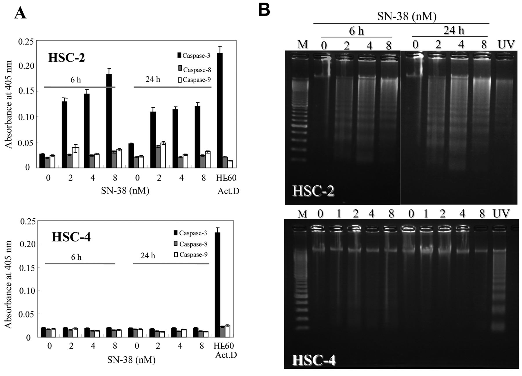
Effect of SN-38 on apoptosis induction in oral squamous cell carcinoma cell lines. HSC-2 and HSC-4 cells were treated with the indicated concentrations of SN-38 for 6 or 24 h, and then assayed for caspase activation (A) and DNA fragmentation (B). Treatment of HL-60 cells with actinomycin D (10 μM) for 2 h was used as a positive control for caspase activation. Each value represents the mean±S.D. from three independent experiments. *p<0.05 (A). DNA prepared from HL-60 cell exposed to UV-irradiation (6 J/m2/min, 1min) followed by 3 h incubation was used as a positive control for DNA fragmentation.
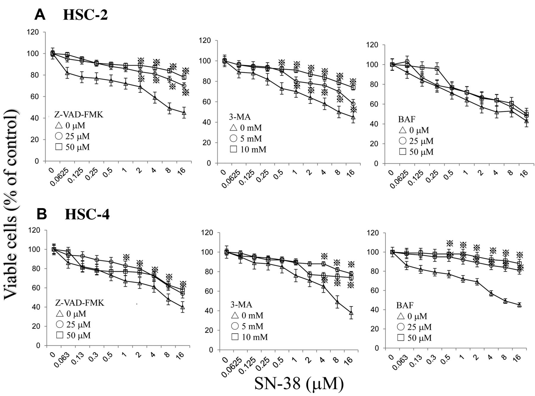
Effect of apoptosis and autophagy inhibitors. In order to investigate the role of apoptosis and autophagy in SN-38-induced cell death, cells were pre-treated with pan-caspase inhibitor (Z-VAD-FMK), or autophagy inhibitors (3-methyladenine, bafilomycin A1) before treatment with SN-38 (Figure 6). 3-Methyladenine is a phosphatidylinositol-3-kinase inhibitor that inhibits the formation of autophagosome (17). Bafilomycin A1 is a V-type proton pump inhibitor that inhibits the formation of secondary lysosome (18). The cytotoxicity of SN-38 towards HSC-2 cells was significantly diminished by pre-treatment with caspase inhibitor, but not with autophagy inhibitors, supporting the involvement of apoptosis in the SN-38-induced cell death of HSC-2 cells. On the other hand, cytotoxicity of SN-38 against HSC-4 cells was significantly diminished by pre-treatment with autophagy inhibitors, but not with caspase inhibitor, supporting the involvement of autophagy in SN-38-induced cell death of HSC-4 cells.
Discussion
The present study demonstrated that SN-38 exhibited much higher cytotoxicity towards OSCC cell lines, as compared with the other four topoisomerase inhibitors. This may be due to its more favorable membrane permeability. The octanol-water partition co-efficient (logP) is a useful marker for evaluating the cytotoxicity of the related compounds. Both membrane permeability and cytotoxicity have been reported to reach maximum levels when the logP value approaches 3 (19, 20). As expected, the logP value of 2.31 for SN-38 is much closer to this value as compared with the other inhibitors (camptotecin: log p=0.95; etoposide: log p=0.28; teniposide: log p=1.2; irinotecan: log p=4.18).

Induction of terminal deoxylnucleotidyl transferase dUTP nick end labeling (TUNEL)-positive cells by SN-38. HSC-2 (A) and HSC-4 cells (B) were seeded onto the chamber slides and cultured for 48 h to achieve complete cell adherence. The cells were then left untreated (control) or treated with SN-38 (8 nM) for 6 or 24 h, and then subjected to TUNEL assay or nuclear staining with propidium iodide (PI) to detect DNA fragmentation and nuclear condensation, respectively. Images were taken by confocal laser scanning microscopy. Original magnification, ×40.

Effect of SN-38 on fine cellular structure of HSC-2 (A) and HSC-4 (B) cells. Cells were treated with SN-38 (8 nM) or without (control) for 12 or 24 h, and fixed for observation under transmission electron microscopy. When the cells were cultured for 12 or 24 h with SN-38, vacuolated mitochondria with electron-lucent matrix (indicated by A) and second lysosome (indicated by arrows in B) were found. Control cells possess healthy mitochondria (indicated by arrow in A). Original magnification, ×2500.
SN-38 was highly cytotoxic in OSCC cells, while it caused little or no damage to oral tissue cells (HGF, HPC and HPLF), yielding quite a high TS value (TS=1321). However, SN-38 did cause significant damage to skin keratinocytes (HEK-a, HEK-n), albeit still maintaining some tumor specificity (TS=22). The higher sensitivity of keratinocytes to SN-38 may be due to their abnormally higher proliferation potential in the hormone and growth factor-enriched medium. It was necessary for us to use this enriched medium, since the keratinocytes cease to grow immediately in the regular culture medium (DMEM supplemented with 10% FBS). This suggests the possibility that SN-38 may target actively proliferating cells. This is supported by our recent observation that the cytotoxicity of SN-38 was significantly augmented by combination with docetaxel or methotrexate which inhibits cell growth time-dependently (unpublished data). Topoisomerase inhibitors such as topotecan, irinotecan and SN-38 have been reported to be substrates for ATP-binding cassette sub-family G member 2 (ABCG2) (6, 7, 21), and the susceptibility of OSCC cells to topoisomerase inhibitors may be explained by lower expression or malfunctioning of transporters such as ABCG2 and P-glycoprotein.
Irinotecan is one of the most popular chemotherapeutic drugs used for the treatment of patients with inoperable colon and breast cancer, with significant life-prolonging effects (22-24). However, irinotecan has not been approved for the treatment of head and neck carcinoma, possibly due to the lack of in vitro data. Considering the highly-specific action of SN-38 towards OSCC cell lines, oral administration of irinotecan to patients with OSCC should be reconsidered.
Other less cytotoxic topoisomerase inhibitors, such as camptothecin and teniposide had a higher tumor specificity towards OCSS vs. oral cells (TS=2961 and 3190, respectively) and keratinocytes (TS=372 and 321, respectively), than SN-38. An appropriate local drug delivery system for these inhibitors for future application to patients with OSCC remains to be explored
The present study also demonstrated that SN-38 induced different types of cell death in two different OSCC cell lines: apoptosis in HSC-2 cells and autophagy in HSC-4 cells, although the sensitivities of these cells to SN-38 are comparable (Table I). Autophagy is also responsible for cell survival under nutritionally-starved conditions (25, 26). The present study suggests that autophagy induced by SN-38 in HSC-4 cells may be responsible for cell death rather than cell survival. The possibility of induction of necrosis seems to be low, considering the absence of any smear pattern of DNA fragmentation (Figure 3B). It is concluded that SN-38 is highly cytotoxic towards OSCC cell lines, regardless of the type of cell death induced.

Effect of caspase and autophagy inhibitors on SN-38-induced cytotoxicity towards OSCC cell lines. HSC-2 (A) and HSC-4 (B) cells were inoculated in 96-well plates and incubated for 48 h to achieve complete adherence. The medium was replaced by fresh medium containing the caspase inhibitor (Z-VAD-FMK), 3-methyladenin (3-MA) or bafilomycin (BAF) and incubated for 60 min. Cells were then treated for 24 h with different concentrations of SN-38 to determine the viable cell number by 3-(4,5-dimethylthiazol-2-yl)-2,5-diphenyltetrazolium bromide method. Each value represents the mean±S.D. from three independent experiments. *p<0.05 compared with untreated controls.
Acknowledgements
This study was supported in part by a Grant-in-Aid from the Ministry of Education, Science, Sports and Culture of Japan (Sakagami, No.19592156). The Authors acknowledge Drs. Kunii and Kanda for their technical assistance.
- Received July 30, 2012.
- Revision received October 8, 2012.
- Accepted October 9, 2012.
- Copyright© 2012 International Institute of Anticancer Research (Dr. John G. Delinassios), All rights reserved
References
In this issue


Jump to section
Related Articles
Cited By...
- Potentiation of Anticancer Activity of G2/M Blockers by Mild Hyperthermia
- Impact of Topoisomerases Complex Deregulation on Head and Neck Carcinoma Genomic Instability
- Cytotoxic Components Against Human Oral Squamous Cell Carcinoma Isolated from Andrographis paniculata
- Search of New Cytotoxic Crude Materials Against Human Oral Squamous Cell Carcinoma Using 1H NMR-based Metabolomics