


Abstract
In this study, combinations of resveratrol with platinum drugs cisplatin and oxaliplatin were administered to human ovarian A2780, A2780cisR and A2780ZD0473R cell lines with the aim of offering a means of overcoming drug resistance. Cell viability was quantified using the 3-(4,5-Dimethyl-2-thiazolyl)-2,5-diphenyl-2H-tetrazolium bromide (MTT) reduction assay. Combination indices and dose response curves were used as measures of the combined drug action. Greatest synergism was observed when resveratrol was administered first followed by the platinum drug (cisplatin or oxaliplatin) 2 h later, and the least synergism was achieved when the two types of compounds were administered as a bolus. The results can be explained by assuming that the administration of resveratrol 2 h before the platinum drug may sensitize the ovarian cancer cells to platinum-induced apoptosis, thus providing a means of overcoming drug resistance. If the results are confirmed in vivo, they may be significant clinically.
With more than 10 million new cases diagnosed every year, cancer is one of the most devastating diseases of our time. It is estimated that by 2030 there would be 27 million cases of cancer and 17 million cancer related deaths annually (1). Although ovarian cancer accounts for only 3% of all types of cancer in women, its management remains a major challenge, being the most lethal gynaecological malignancy and a cause of considerable morbidity (2).
Platinum drugs such as cisplatin and oxaliplatin (Figure 1) are routinely used to treat various types of cancer including the ovarian cancer. Unfortunately, almost all relapsing ovarian cancers eventually develop platinum resistance; patients with resistant tumours have a median survival time of 6 months, with only 27% living longer than 12 months (3). The development of chemoresistence and the recurrence of secondary tumours serve to put chemotherapy on the back foot in the fight against cancer. Phytochemicals provide a natural source of drugs for the treatment of various diseases including cancer. One such phytochemical is resveratrol, which is 3,5,4’-trihydroxystilben (Figure 1). Found in red grapes, mulberries and some nuts, resveratrol has potential antitumourigenic and anti-inflammatory activities. Because of its broad-spectrum health benefits, resveratrol has been termed as the nature's state-of-the-art medicine. As applied to cancer, reseveratrol has both chemopreventive and chemotherapeutic potentials. Whereas chemoreventive action may be associated with blocking carcinogen activation, boosting antioxidant capacity and arresting cell proliferation, chemotherapy may be associated with inducing apoptosis of damaged/transformed cells, turning off the angiogenic switches, blocking neovascularization in tumour cells, suppressing invasion and metstasis, and sensitizing tumour cells to undergo chemotherapy-induced apoptosis (4). In a pioneering study, Jang and co-workers reported that resveratrol was effective in blocking all stages of carcinogenesis including initiation, promotion and progression (5).
The antitumour activity of resveratrol is believed to be due to inhibition of cancer cells proliferation and induction of apoptosis. The major factors involved in the development of drug resistance are multidrug resistance (MDR) gene, nuclear transcription factor-kB (NF-kB), and serine/threonine protein kinase AKT. About 15% of all solid tumours are driven by NF-kB as a player whereas most cancer preventive agents act as NF-kB inhibitors (6). Aberrant activation of NF-kB that may occur in response to a variety of stimuli, such as cytokines, growth factors, physiological, physical and oxidative stress, and certain pharmacological drugs and chemicals (7), can provide malignant cells with protection against apoptosis and stimulate their proliferation. In addition, over-expression of NF-kB is causally linked to phenotypic changes that are characteristic of neoplastic transformation. One way resveratrol overcomes chemoresistance is through the down-regulation of NF-kB. It also induces apoptosis by down-regulating signal transducer and activator transcription factor 3 (STAT3), anti-apoptotic and cell survival gene products (8), such as BCL-2 (9), and also by regulating cyclooxygenase expression (10). Up-regulation of the tumour suppressor p53 and cytokine (MIC-1) that possesses anti-tumourigenic activity are also caused by resveratrol (11). Under anaerobic conditions, Res also acts as a radiation-protecting agent (12). Resveratrol also increases the expression and/or activity of glutathione S-transferase (GST) and glutathione peroxidase (GPx), resulting in lowering of the glutathione level (8). It is thus logical to assume that platinum drugs in combination with reseveratrol may cause enhanced cell kill due to synergistic action and/or compensation of the adverse effects. Such combination treatment may also result in a decrease in systemic toxicity caused by chemotherapies or radiotherapies because of lower doses required (13).

Structures of a) cisplatin, b) oxaliplatin, and c) resveratrol.
The present study aimed to investigate the effect on the cell kill in the human ovarian A2780 (parent), A2780CisR (cisplatin-resistant type) and A2780ZD0473R (ZD0473-resistant type) cancer cells due to the combination of cisplatin and oxaliplatin with resveratrol (Figure 1). It constitutes a part of our continued study on synergism from combinations of platinum drugs with phytochemicals (14-16).
Materials and Methods
Oxaliplatin and resveratrol in powder form were obtained from Sigma Aldrich, Sydney Australia and cisplatin was prepared according to the method of Dhara (17). The compounds were initially dissolved in dimethylformamide (DMF) followed by the addition of milli Q (mQ) water (at a ratio 1:5) to give 1 mM stock solutions. The solutions were filtered using DISMIC-25cs ADVANTEC filter (cellulose acetate, 0.20 μm hydrophilic, pressure limitation: 0.51 MPa) to sterilize. The stock solutions were serially diluted with freshly prepared RPMI-1640 medium to produce a range of final concentrations from 0.0005 to 100 μM. A2780, A2780cisR and A2780ZD0473R ovarian cancer cell lines were obtained from Ms. Zhang from the Royal Prince Alfred Hospital, The University of Sydney, Australia. The cell lines were sub-cultured in RPMI 1640 medium that was prepared in 10% fetal calf serum (FCS, 1 mM Hepes, 5.6% sodium bicarbonate and 200 mM glutamine.
(IC50) values (μM) of cisplatin, oxaliplatin and resveratrol for the human ovarian cancer A2780, A2780cisR and A2780ZD0473R cell lines.
Cytotoxicity assays. The cell killing effect was determined using the 3-(4,5-Dimethyl-2-thiazolyl)-2,5-diphenyl-2H-tetrazolium bromide (MTT) reduction assay (18). Briefly, 4000 to 5000 cells per well in RPMI-1640 medium were seeded into flat-bottomed 96-well culture plate and allowed to attach overnight. For single treatments, drugs were added at a range of at least three to five different concentrations to triplicate wells which were left in the incubator (37°C, 5% carbon dioxide in air, pH7.4) for 72 h. After preparation of serial five-fold dilutions of the drugs in 10% FCS/RPMI 1640 medium (for oxalilatin: 0.064-8 μM, for cisplatin: 0.32-40 μM, and for resveratrol: 1.6-200 μM), 100 μl of drugs were added to equal volumes of cell culture in triplicate wells and cells were then left to incubate under normal growth conditions for 72 h at 37°C in a humidified atmosphere. For combination studies, cells were treated with increasing concentrations of compounds at constant ratios of their IC50 values using the sequences: 0/0, 0/2 and 2/0, where 0/0 meant that both the drugs were added at the same time, 0/2 meant that Cis or Oxa was added first followed by the Res 2 h later and 2/0 meant the converse. The concentration ranges were: cisplatin: 0.26-4.09 μM, 1.66-26.52 μM and 0.57-9.11 μM; oxaliplatin: 0.16-2.62 μM, 0.59-9.41 μM and 0.42-6.75 μM; resveratrol: 9.91-158.57 μM, 10.66-170.54 μM and 9.93-158.86 μM for A2780, A2780CisR and A2780ZD0473R cell lines, respectively. At the completion of the 72 h period of incubation, the MTT assay was performed as in previous experiments (18).
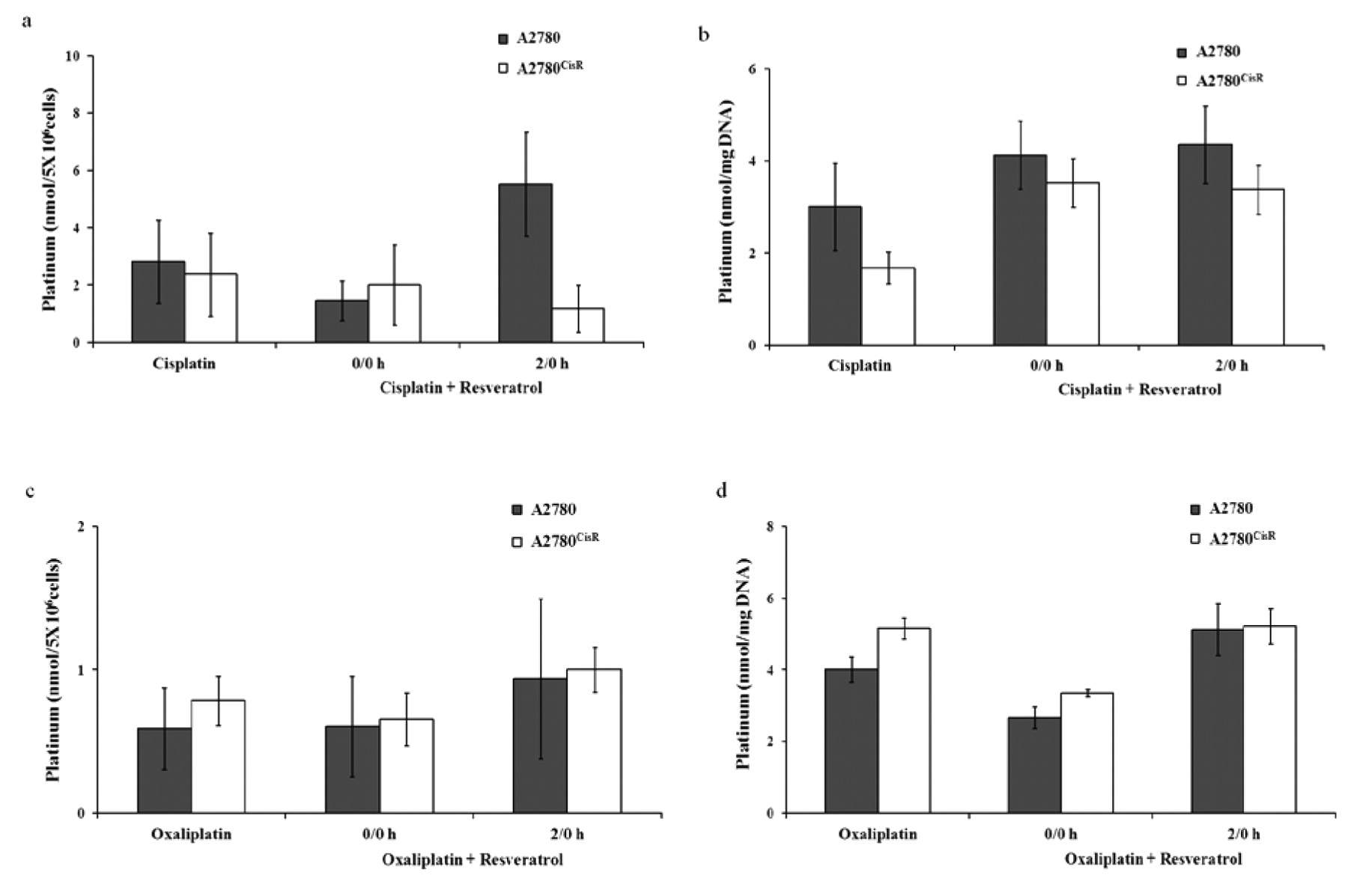
a) Total intracellular platinum levels and b) platinum–DNA binding levels from cisplatin alone and its 0/0 h and 2/0 h combination with resvertraol in the A2780 and A2780cisR cell lines, c) total intracellular platinum levels and d) platinum–DNA binding levels from oxaliplatin alone and its 0/0 h and 2/0 h combination with resveratrol in the A2780 and A2780cisR cell lines.
Dose-effect parameters applying to combinations of cisplatin and oxaliplatin with resveratrol in the A2780, A2780cisR and A2780ZD0473R cell lines.

Electrostatic potential map for (DFT) optimized structure of resveratrol.
The combined action of the drugs was studied using the median effect analysis. Combination index (CI) was calculated based on the pooled data from five individual experiments each comprising at least three data points for each drug alone and for the drug combinations. The CI for two drugs in combination can be calculated using the following equation (19, 20).
 where D1 and D2 represent the concentrations of compounds 1 and 2 in combination to achieve x% inhibition whereas D1x and D2x represent concentrations of compounds 1 and 2 to achieve x% inhibition when present alone. Dx can be readily calculated from the following equation, where Dx denotes dose of drug, Dm is the median-effect dose or IC50, fa is the fraction of cells affected (killed) by the dose, fu is the fraction of cells remaining unaffected so that fu=1-fa and m is the exponent defining the shape of the dose effect curve.
 The CI of <1, =1 and >1 indicates synergism, additiveness and antagonism in the combined drug action respectively. The CI, Dm and linear correlation coefficient r values were calculated using Calcusyn software (V2) (Biosoft, UK). The r value indicates the goodness of fit for the pooled data (where r=1 is a perfect fit) and r of median effect plot for the cell culture system should be greater than 0.95 (19, 20).
Platinum accumulation and platinum–DNA binding. Cellular accumulation of platinum and platinum–DNA binding levels were measured in order to determine whether the values were affected by the sequence of addition. It is believed that the results may aid in the understanding of the combined drug action. The most synergistic (2/0) and least synergistic (0/0) combinations of cisplatin and oxaliplatin with resveratrol were selected for the determination of cellular platinum accumulation and platinum–DNA binding level. The method used for the determination of total intracellular platinum and platinum–DNA level was a modification of that described by Di Blasi et al. (21). Platinum drugs and resveratrol were added to culture plates containing exponentially growing A2780 and A2780cisR cells in 5 ml 10% FCS/RPMI 1640 culture medium (cell density=1×10⁶ cells ml−1 and cells were incubated for 24 h, at the end of which cell monolayers were trypsinized and cell suspension (5 ml) was transferred to centrifuge tube and spun at 3500 rpm for 2 min at 4°C. After washing twice with ice-cold phosphate-buffered saline (PBS) cell pellets were stored at −20°C until assayed.
Cellular accumulation. Cell pellets from drug combinations were suspended in 0.5 ml 1% triton-X, held on ice while being sonicated. Total intracellular platinum contents were determined by graphite furnace (AAS) using a Varian SpectrAA-240 plus with GTA 120 atomic absorption spectrophotometer (Varian Australia Pty Ltd Melbourne Victoria Australia) (22).
Platinum–DNA binding. After drug incubation, DNA was isolated from cell pellet using H440050 JETQUICK Blood DNA Spin Kit/50 (Austral Scientific Pty Ltd Sydney NSW Australia) according to the modified protocol of Bowtell (23). DNA content was determined by UV spectrophotometry (260 nm) (Varian Cary 1E UV-Visible with Varian Cary Temperature Controller) and platinum levels was determined by graphite furnace AAS. A260/A280 ratios were found to be between 1.75 and 1.8 for all samples, reflecting high sample purity (24). The DNA concentration was calculated according to the equation: Concentration=absorbance at 260 nm × 50 ng/μl.
Results
Growth-inhibitory effect of single drugs. Table I gives the IC50 values and resistant factors (RF) for cisplatin, oxaliplatin and resveratrol for the ovarian A2780, A2780cisR and A2780ZD0473R cancer cell lines. RF is defined as the ratio of concentration of the drug required for 50% cell kill in the resistant cell line to that in the parent cell line. Among three compounds, oxaliplatin was found to be most active and Res the least. However, resveratrol gave much lower RFs than the platinum drugs.
Growth-inhibitory effects of drugs in combination. CIs were used as a measure of combined drug action. As stated earlier the main objective of the present study was to investigate synergism in activity from the combinations of the selected platinum compounds with the phytochemical Res in three human ovarian cancer cell lines. Table II gives dose–effect parameters in terms of median-effect dose, shape (sigmoidicity), conformity (linear correlation coefficient), represented as Dm, m and r respectively. The CIs indicate that administration of resveratrol and platinum drug with a 2 h time gap produced much greater cell kill than the bolus administration and that the combined drug action was greater when resveratrol was added first than the converse. The degree of synergism was greater for the combination of resveratrol with cisplatin than for its combination with oxaliplatin.
Cellular platinum accumulation and platinum–DNA binding levels. Figure 2 gives the cellular accumulation of platinum and platinum–DNA binding levels in A2780 and A2780cisR cell lines applying to the 0/0 h and 2/0 h combinations of cisplatin and oxaliplatin with reseveratrol It was found that in both A2780 and A2780cisR cell lines, the cellular accumulation of platinum and platinum–DNA binding level resulting from the 2/0 combination were significantly greater (more so for the accumulation than the binding level) than that resulting from the 0/0 combination and the equivalent concentration of cisplatin or oxaliplatin present alone. As applied to the combination of oxaliplatin with resveratrol in A2780 and A2780cisR cell lines, cellular platinum and platinum–DNA binding level were greater in the resistant cell line than in the parent cell line whereas the converse was true for the combination of cisplatin with resveratrol.
Discussion
In this study, we investigated sequence-dependence of the combined drug action for the binary combinations of cisplatin and oxaliplatin with the phyochemical resveratrol. It was found that the administration of resveratrol 2 h before that of the platinum drug produced much greater cell kill than the converse and the bolus as it was reported to be the case in the combination of cisplatin and oxaliplatin with quercetin and thymoquinone (16). Much greater synergism resulting from the 2/0 combination of the platinum drugs with resveratrol, quercetin and thymoquinone indicates that the incubation of the ovarian cancer cells with the phytochemicals for a short period before the administration of the platinum drug served to sensitize them for platinum-induced cell death. The enhanced cell kill due to combination of resveratrol with cisplatin and Ooxaliplatin cannot be attributed simply to the antioxidant role played by the polyphenol because when resveratrol acts as an antioxidant, it would serve to reduce oxidative stress, resulting in sparing of the reduced form of glutathione (GSH). An increased concentration of GSH would cause increased deactivation of the platinum drug before its binding with the DNA. The increased platinum–DNA binding level observed for the 2/0 sequence of administration clearly indicates that the above explanation cannot be true. As noted earlier, resveratrol increases the transcription of phase II enzyme GPx resulting in a lower GSH level (8). The resveratrol-induced lowering of GSH level can be seen to provide an explanation for the increased platinum–DNA binding level due to prior incubation of ovarian cancer cells with Res. Another reason for the increased platinum–DNA binding level is due to the inhibition of P-glycoprotein function by reseveratrol (25). P-Glycoprotein is considered to be a key player in the development of chemoresistance as it reduces concentrations of chemotherapeutic drugs such as cisplatin and oxaliplatin within tumour and hence serves to lower their efficacy. By inhibiting P-glycoprotein function, resveratrol increases the accumulation of cisplatin and oxaliplatin and hence their efficacy as well.
Resveratrol targets many components of intracellular signaling pathways including pro-inflammatory mediators, regulators of cell survival and programmed cell death, and tumour angiogenic and metstastic switches (4). It does so by modulating a distinct set of upstream kinases, transcription factors and the associated regulators. The antiproliferative and growth inhibitory effect of resveratrol have been attributed to its ability to block DNA synthesis and interfere with various stages of cell cycle progression. Multiple lines of evidence indicate that resveratrol induces cell death by activating pro-apoptotic signaling molecules, as well as by inhibiting anti-apoptotic molecules of intracellular signal transduction pathways. According to Kundu and Suhr (4), upstream kinases and transcription factors in the intracellular network are the potential targets of resveratrol in normal and cancerous cells. These signal transducers are fine tuned in normal cells, while many of them are deregulated in cancerous cells. Depending on the cell type and stimulus, resveratrol may either suppress the aberrant activation of particular signaling pathways or restore activities of others whose function is suppressed. Both the platinum resistance and the enhancement of platinum action due to combination of cisplatin and oxaliplatin with resveratrol also appear to be related to NF-kB. Whereas the resistance to platinum drugs is associated with aberrant activation of NF-kB, resveratrol is known to dampen its expression at higher concentration (>20 μM) but enhance its activation at a lower concentration range (1-10 μM). Although only about 15% of all solid tumours are driven by NF-KB as a player, most cancer–preventive agents are believed to be NF-kB inhibitors (6). We suggest that prior incubation of ovarian cancer cells with resveratrol serves to reduce the expression of NF-kB and hence lower the resistance to the platinum drugs. Proteomic studies designed to characterize key proteins that are differentially expressed in resistant cell lines as compared to the parent cell line and whose functions are restored to normalcy due to treatment with successful drug combinations may provide further light on this matter.
We carried out optimization of the structure of resveratrol at the DFT level (B3LYP/6-31G*) following molecular mechanics and semi-empirical optimizations using the program Spartan ’10 V1.1.0 (Spartan ’10 Version 1.1.0 Wavefunction, Inc.) in order to obtain molecular parameters of the compound. It was thought the calculated values might be useful in explaining its biological functions. It was found that the electrostatic potential map (Figure 3) has significant electron-rich regions, in line with the antioxidant role played by resveratrol. Absence of any significant electron-deficient region indicates that the phytochemical may not be damaging to biomolecules such as DNA and proteins. The (LUMO-HOMO) energy difference of 389.3 kJ/mol (4.03 eV) indicates that resveratrol would be neither extremely labile nor extremely inert kinetically. The calculated dipole moment of Res is 0.68 debye and its logP is 3.06, indicating that its lipid solubility would be significant. Other (QSAR) parameters of the compound are: area=256.92 Å2 (with accessible area being 195.44 Å2), volume=236.85 Å3 and polarizability=59.63.
Acknowledgements
Meher Un Nessa is grateful to the Australian Department of Education, Employment and Workplace Relations (DEEWR) for the Endeavour Postgraduate Award and Sydney Medical School, University of Sydney for Part-fee scholarship. Meher Un Nessa is also grateful to Khulna University, Bangladesh for providing study leave to carry out the study in the University of Sydney, Australia. This work is partly funded by Biomedical Science Research Initiative Grant and Biomedical Science Cancer Research Donation Fund.
Footnotes
-
Conflict of Interest
Meher Un Nessa, Philip Beale, Charles Chan, Jun Qing Yu and Fazlul Huq declare that they have no financial and personal relationships with other people or organizations that could inappropriately influence their work.
- Received October 10, 2011.
- Revision received December 8, 2011.
- Accepted December 9, 2011.
- Copyright© 2012 International Institute of Anticancer Research (Dr. John G. Delinassios), All rights reserved


