
Main menu

  • Home
  • Current Issue
  • Archive
  • Info for
    • Authors
    • Editorial Policies
    • Subscribers
    • Advertisers
    • Editorial Board
    • Special Issues 2025
  • Journal Metrics
  • Other Publications
    • In Vivo
    • Cancer Genomics & Proteomics
    • Cancer Diagnosis & Prognosis
  • More
    • IIAR
    • Conferences
    • 2008 Nobel Laureates
  • About Us
    • General Policy
    • Contact
  • Other Publications
    • Anticancer Research
    • In Vivo
    • Cancer Genomics & Proteomics

User menu

  • Register
  • Subscribe
  • My alerts
  • Log in
  • My Cart

Search

  • Advanced search
Anticancer Research
  • Other Publications
    • Anticancer Research
    • In Vivo
    • Cancer Genomics & Proteomics
  • Register
  • Subscribe
  • My alerts
  • Log in
  • My Cart
Anticancer Research

Advanced Search

  • Home
  • Current Issue
  • Archive
  • Info for
    • Authors
    • Editorial Policies
    • Subscribers
    • Advertisers
    • Editorial Board
    • Special Issues 2025
  • Journal Metrics
  • Other Publications
    • In Vivo
    • Cancer Genomics & Proteomics
    • Cancer Diagnosis & Prognosis
  • More
    • IIAR
    • Conferences
    • 2008 Nobel Laureates
  • About Us
    • General Policy
    • Contact
  • Visit us on Facebook
  • Follow us on Linkedin
Case ReportClinical Studies

Dermatofibrosarcoma Protuberans: A Case Series of 16 Patients Treated in a Single Institution with Literature Review

MARIA ARCHONTAKI, DIMITRIS P. KORKOLIS, NIKI ARNOGIANNAKI, CHARIKLEIA KONSTANTINIDOU, SPYROS GEORGOPOULOS, PANAGIOTIS DENDRINOS, GEORGIOS ZARKADAS and GEORGIOS KOKKALIS
Anticancer Research September 2010, 30 (9) 3775-3779;
MARIA ARCHONTAKI
  • Find this author on Google Scholar
  • Find this author on PubMed
  • Search for this author on this site
  • For correspondence: marchontaki@web.de
DIMITRIS P. KORKOLIS
  • Find this author on Google Scholar
  • Find this author on PubMed
  • Search for this author on this site
NIKI ARNOGIANNAKI
  • Find this author on Google Scholar
  • Find this author on PubMed
  • Search for this author on this site
CHARIKLEIA KONSTANTINIDOU
  • Find this author on Google Scholar
  • Find this author on PubMed
  • Search for this author on this site
SPYROS GEORGOPOULOS
  • Find this author on Google Scholar
  • Find this author on PubMed
  • Search for this author on this site
PANAGIOTIS DENDRINOS
  • Find this author on Google Scholar
  • Find this author on PubMed
  • Search for this author on this site
GEORGIOS ZARKADAS
  • Find this author on Google Scholar
  • Find this author on PubMed
  • Search for this author on this site
GEORGIOS KOKKALIS
  • Find this author on Google Scholar
  • Find this author on PubMed
  • Search for this author on this site
  • Article
  • Figures & Data
  • Info & Metrics
  • PDF
Loading

Abstract

Background: Dermatofibrosarcoma protuberans is an uncommon skin tumour with a low to intermediate-grade of malignancy, characterized by progressive growth and a propensity for local recurrence. Patients and Methods: A retrospective study of a series of 16 consecutive patients with dermatofibrosarcoma protuberans who were treated in the host Institution over the last seven years was performed, with special emphasis on the outcome and disease-free interval, as well as recurrence rate over a mean follow-up period of 43.65 months. Results: The clinicopathological features and results were reviewed. The primary treatment consisted of wide local excision with or without radiotherapy on 13 patients with primary and 3 with recurrent disease, and all patients remained free of disease recurrence during the mean follow-up period. Conclusion: The results of this study and a review of the literature support the notion that aggressive, wide surgical resection with disease-free margins, with or without radiotherapy decreases local recurrences and offers an excellent probability of cure. The accumulated data also confirm that all patients with dermatofibrosarcoma protuberans should be followed up for an extended period, beyond the usual recommended 5-year follow-up, because late recurrences may occur.

  • Dermatofibrosarcoma protuberans
  • surgical treatment
  • tumour
  • wide local excision

Dermatofibrosarcoma protuberans (DFSP) is a rare cutaneous soft tissue sarcoma that was first described by Taylor in 1890 (1, 2), clinically was classified by Darier and Ferrand (3) and later named by Hoffman (4). It is a rare skin tumour that clinically often masquerades as a benign, indolent tumour, but microscopically it extends far beyond assessed clinically margins, and spreads locally in the dermis, subcutaneous tissue and muscles (5). Approximately 85-90% of all DFSPs are low-grade lesions while the rest contain a high grade fibrosarcomatous component and are considered to be intermediate-grade neoplasms (6).

The goal of this retrospective review of 16 patients who underwent wide local excision with or without radiotherapy for the treatment of DFSP tumours was to expand on the clinicopathological characterization and treatment strategy of this unusual neoplasm. Awareness of this entity is important for a prompt diagnosis and a proper management of the disease, preventing both over- and undertreatment of this low to intermediate-grade malignancy.

Materials and Methods

A retrospective chart review was performed on all patients with a history of DFSP who were treated in the host Institution from November 2001 to October 2009. All patients that were still alive were contacted for current performance status. The diagnosis of DFSP was established on 16 patients histopathologically by H&E staining and with positive CD34 immunohistochemical markers. Surgical treatment consisted of wide local excision with at least a 3-cm margin, including the underlying fascia. Adjuvant radiation therapy was given to patients with large lesions or close margins after maximal resection. The details of the radiation therapy were not analysed. A review of the medical records provided information on demographics (age, sex), tumour characteristics (location, size), treatment course, evolution (location and time of recurrence) and length of follow-up.

Results

From November 2001 to October 2009, 16 patients were diagnosed with DFSP.

A summary of the clinical data is presented in Table I. The mean patient age at time of diagnosis was 41.12 years (range, 21-59 years). Preoperative duration of symptoms was described by all patients to be long (more than 1 year). Sex distribution (9 females, 7 males) was approximately equal. The anatomical location of the tumour was on the trunk and proximal extremities in 9, on the lower extremity (Figures 1 and 2) in 2 and on the head and neck in 5. Tumour size ranged from 1 to 22.5 cm (median 3.32 cm). The majority presented clinically as a raised slowly growing firm, nodular dermal/subcutaneous mass. The median follow-up in this series was 43.65 months.

View this table:
  • View inline
  • View popup
  • Download powerpoint
Table I.

Clinical data.

A history of resection and subsequent local recurrence was found in 3 patients and all were associated with positive or very close initial surgical margins. No case gave rise to metastatic disease. Treatment in all cases was wide local excision with disease-free margins of at least 2.5 cm.

Reconstruction of the defects took place primarily in 9 cases and with split-thickness skin grafts in three patients. One patient underwent reconstruction with a pedicled Latissimus dorsi flap, while one another with a pedicled trapezius flap. In addition, a free radial forearm flap was used as a reconstructive surgical procedure in two patients. Adjuvant radiotherapy was provided in two cases with local recurrence and in one case of massively extended disease.

Discussion

DFSP is a rare, low- to intermediate-grade mesenchymal malignant neoplasm that originates from the dermis, from which it often invades the subcutaneous tissue (7, 8), and accounts for 1% to 2% of all soft tissue sarcomas (9, 10). DFSP is characterized by an indolent, very slow but infiltrative growth pattern and in many cases its symptoms are long lasting, up to 30 years according to Lindner et al. (11). Preceding traumas are mentioned in 10-20% of cases and some tumours are found in burn and surgical scars (12). DFSPs arise as pink or violet-red plaques, while the surrounding skin may be telangiectatic (13, 14). They typically do not exhibit a nodular growth pattern until late in their course (15). Small lesions are mobile but as they enlarge they become fixed because of infiltration of the underlying fascia (13, 14). Clinically, the largest diameter in the majority of DFSPs measures less than 5 cm. The plaque stage may clinically cause confusion with scar tissue or sclerosing dermatoses such as morphea or scleroderma (16). Intermediate lesions can be confused with larger dermatofibromas while advanced lesions must be differentiated from malignant fibrous histiocytoma and fibrosarcoma (17).

DFSP typically arises on the trunk of the body and proximal extremities, followed by the distal extremities, and then head and neck with a slight male predomination (18). It usually presents in the fourth decade of life, although the age of appearance varies widely (19, 20). In the current series, the tumours had a similar age distribution to typical DFSP, with a slight female preponderance. The most common sites of presentation for DFSP in the current series were the trunk, followed by the head and neck and then extremities, but these apparent differences may reflect sampling error in this relatively small case series.

Uncommon variants of DFSP include the Bednar tumour (pigmented DFSP), myxoid, atrophic, fibrosarcomatoid, mixed granular cell and sclerosing. A congenital variant has also been described with characteristic histological markers (7, 21, 22).

Figure 1.
  • Download figure
  • Open in new tab
  • Download powerpoint
Figure 1.

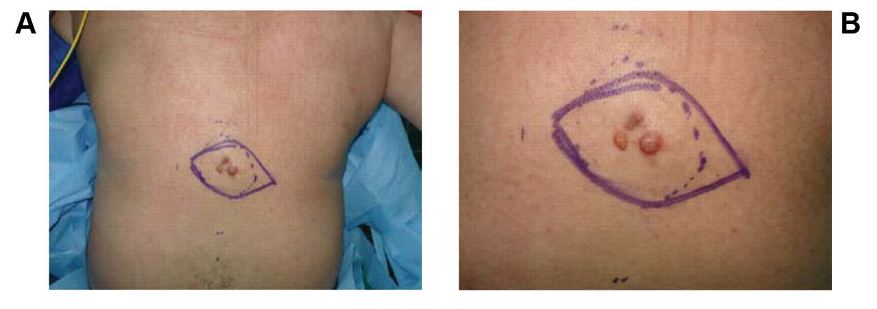
Case 8, DFSP on the back with multinodular protuberant ‘phenotype’.

Figure 2.
  • Download figure
  • Open in new tab
  • Download powerpoint
Figure 2.

A: Case, 16 DFSP on the thigh with nodular appearance. B: Case 7, DFSP on the anterior thoracic wall with plaque-like appearance.

Figure 3.
  • Download figure
  • Open in new tab
  • Download powerpoint
Figure 3.

A: The tumor was stained positively for CD34 (×400). B: Storiform pattern infiltrating between adnexal structures. Fat is also involved by tumor cells (HE,×100).

Histologically DFSP is composed of monomorphous spindle-shaped cells arranged in a storiform pattern on a background of fibrous stroma. A honeycomb pattern of infiltration into the subcutaneous fat is also seen, while mitoses are rare, explaining the low grade malignant behavior of the tumour (6, 23). DFSP is strongly positive to staining with CD34. It is also immunoreactive to vimentin. On the contrary, it shows a light or absent reaction to CD44, which represents a useful marker for the differential diagnosis from dermatofibroma. Distinctive histological features of DFSP, as well as findings consistent with the immunohistochemichal profile of typical DFSP were present in all the cases in this series (Figure 3).

More than 90% of DFSP are characterized by a reciprocal chromosomal translocation, t(17;22)(q22;q13), or more frequently, a supernumerary ring chromosome composed of hybrid material derived from t(17;22). This rearrangement leads to constitutive activation of the platelet-derived growth factor as a result of deregulated ligand expression (24).

The main feature of DFSP is the horizontal spread of the tumour by creating neoplastic projections to all directions, like pseudopodia, resulting in the ejection of neoplastic cells up to 3 cm peripherally to the main tumour. This histological behaviour results in the high recurrence rate, as a result of satellite neoplastic cell populations that may be left in situ, during resection (13, 25).

According to Gayner et al. (26), excisional microscopic negative border less than 2 cm represents the main predisposing factor of recurrence. Bowne et al. (25) established as negative prognostic factors for recurrence of DFSP the age older than 50 years, resection close to positive microscopic margins, fibrosarcomatous variant, high mitotic rates and increased cellularity. In addition, it is also well accepted that the number of recurrences up to the last excision represents a negative prognostic factor for the possibility of future recurrence.

DFSP has a tendency for local recurrence of up to 25% and most recurrences tend to develop within 3 years, after resection. Late recurrences, even after 15 years, have been also reported (18, 27, 28). Metastasis may occur through the bloodstream to the lungs or less commonly via the lymphatics to regional lymph nodes (29, 30). Recent study by Fiore et al. (15) confirms that most cases of DFSP, including recurrent disease, can be managed with wide-excision surgery, providing a crude incidence of recurrence in the entire population 3% at 5 years and 4.2% at 10 years and a crude incidence of distant metastasis of 1.7% at both 5 and 10 years. The prognosis of DFSP is generally excellent if completely excised with clear margins (15).

In the current series, no patient relapsed within the mean follow-up time of 43.65 months. On the other hand however, in 2 cases there was a history of excision and recurrence after more than 10 years.

Excision of DFSP is usually performed at the stage of solitary nodulous growth. Because of its harmless general appearance, an inadequate excision of microscopic extensions which are located lateral to the tumour or deeper than it, may take place (12). The pattern of growth which is primarily lateral or radial spread may become deep or vertical in longstanding or recurrent tumours. The first resection is of major importance, since tumour spread after an inadequate first resection may lead to uncontrollable local growth (10).

Most authors suggest a margin of 2.5 to 3 cm of normal tissue from the gross tumour boundary, with a three-dimensional resection (en bloc removal) that includes skin, subcutaneous tissue and the underlying investing fascia, achieving negative microscopic margins (15, 31, 32). An alternative option represents Mohs micrographic surgery, designed for tumours with extensive subclinical skin involvement. Recurrence rates with this surgical potentially tissue-sparing option of 0% to 6.6% have been reported in several series (5, 19, 30). DuBay et al. (20) introduced a multidisciplinary combination approach with DFSP as an efficacious treatment strategy, in order to optimize patient care.

DFSP is considered to be a radiosensitive tumour. Consequently, radiation therapy can be recommended as adjuvant therapy in cases with positive margins or close margins after maximal resection, in case of a large lesion excised with negative margins, in case of recurrent lesion or in case that achievement of wide margins would result in a functional or cosmetic deficit (1, 24, 33).

Recently, imatinib a potent selective inhibitor of the platelet-derived growth factor receptor has been reported to induce complete or partial remissions in most patients with advanced DFSP, and is approved for treatment of adult patients with unresectable, recurrent and/or metastatic DFSP who are not eligible for surgery (34).

In summary, DFSP is a distinct oncological entity which often presents a diagnostic challenge. The proper identification and diagnosis is clinically important. The cornerstone of DFSP management consists of complete surgical resection with negative margins (26). The Authors' experience recommends as a frontline treatment wide excision of the tumour with at least 5 cm of surrounding tissue, extending deeply to histopathological disease-free anatomic tissue, in order to improve prognosis over time. In addition, the authors recommend close follow-up for cancer surveillance for an extended period, as late recurrences may occur.

  • Received April 20, 2010.
  • Revision received May 22, 2010.
  • Accepted May 28, 2010.
  • Copyright© 2010 International Institute of Anticancer Research (Dr. John G. Delinassios), All rights reserved

References

  1. ↵
    1. Suit H,
    2. Spiro I,
    3. Mankin HJ,
    4. Efird J,
    5. Rosenberg AE
    : Radiation in management of patients with dermatofibrosarcoma protuberans. J Clin Oncol 14(8): 2365-2369, 1996.
    OpenUrlAbstract
  2. ↵
    1. Taylor RW
    : Sarcomatous tumours resembling in some respects keloid. J Cutan Genitourin Dis 8: 384-387, 1890.
    OpenUrl
  3. ↵
    1. Darier J,
    2. Ferrand M
    : Dermatofibromes progressifs et residivants on fibrosarcomes de la peau. Ann Dermatol Syphilol 5: 545-570, 1924.
    OpenUrl
  4. ↵
    1. Arnaud EJ,
    2. Perrault M,
    3. Revol M,
    4. Servant JM,
    5. Banzet P
    : Surgical treatment of dermatofibrosarcoma protuberans. Plast Reconstr Surg 100(4): 884-895, 1997.
    OpenUrlCrossRefPubMed
  5. ↵
    1. Snow SN,
    2. Gordon EM,
    3. Larson PO,
    4. Bagheri MM,
    5. Bentz ML,
    6. Sable DB
    : Dermatofibrosarcoma protuberans: a report on 29 patients treated by Mohs micrographic surgery with long-term follow-up and review of the literature. Cancer 101(1): 28-38, 2004.
    OpenUrlCrossRefPubMed
  6. ↵
    1. Mentzel T,
    2. Beham A,
    3. Katenkamp D,
    4. Dei Tos AP,
    5. Fletcher CD
    : Fibrosarcomatous (‘high-grade’) dermatofibrosarcoma protuberans: clinicopathologic and immunohistochemical study of a series of 41 cases with emphasis on prognostic significance. Am J Surg Pathol 22(5): 576-587, 1998.
    OpenUrlCrossRefPubMed
  7. ↵
    1. Bergin P,
    2. Rezaei S,
    3. Lau Q,
    4. Coucher J
    : Dermatofibrosarcoma protuberans, magnetic resonance imaging and pathological correlation. Australas Radiol 51: B64-66, 2007.
    OpenUrlCrossRefPubMed
  8. ↵
    1. Breuninger H,
    2. Sebastian G,
    3. Garbe C
    : Dermatofibrosarcoma protuberans-an update. J Dtsch Dermatol Ges 2(8): 661-667, 2004.
    OpenUrlPubMed
  9. ↵
    1. Greco A,
    2. Roccato E,
    3. Miranda C,
    4. Cleris L,
    5. Formelli F,
    6. Pierotti MA
    : Growth-inhibitory effect of STI571 on cells transformed by the COL1A1/PDGFB rearrangement. Int J Cancer 92(3): 354-360, 2001.
    OpenUrlCrossRefPubMed
  10. ↵
    1. McPeak CJ,
    2. Cruz T,
    3. Nicastri AD
    : Dermatofibrosarcoma protuberans: an analysis of 86 cases-five with metastasis. Ann Surg 166(5): 803-816, 1967.
    OpenUrlPubMed
  11. ↵
    1. Lindner NJ,
    2. Scarborough MT,
    3. Powell GJ,
    4. Spanier S,
    5. Enneking WF
    : Revision surgery in dermatofibrosarcoma protuberans of the trunk and extremities. Eur J Surg Oncol 25(4): 392-397, 1999.
    OpenUrlCrossRefPubMed
  12. ↵
    1. Smola MG,
    2. Soyer HP,
    3. Scharnagl E
    : Surgical treatment of dermatofibrosarcoma protuberans. A retrospective study of 20 cases with review of literature. Eur J Surg Oncol 17(5): 447-453, 1991.
    OpenUrlPubMed
  13. ↵
    1. Maggoudi D,
    2. Vahtsevanos K,
    3. Psomaderis K,
    4. Kiesaridou D,
    5. Valery R,
    6. Karakinaris G
    : Dermatofibrosarcoma protuberans of the face: report of 2 cases and an overview of the recent literature. J Oral Maxillofac Surg 64(1): 140-144, 2006.
    OpenUrlCrossRefPubMed
  14. ↵
    1. Mendenhall WM,
    2. Zlotecki RA,
    3. Scarborough MT
    : Dermatofibrosarcoma protuberans. Cancer 101(11): 2503-2508, 2004.
    OpenUrlCrossRefPubMed
  15. ↵
    1. Fiore M,
    2. Miceli R,
    3. Mussi C,
    4. Lo Vullo S,
    5. Mariani L,
    6. Lozza L,
    7. Collini P,
    8. Olmi P,
    9. Casali PG,
    10. Gronchi A
    : Dermatofibrosarcoma protuberans treated at a single institution: a surgical disease with a high cure rate. J Clin Oncol 23(30): 7669-7675, 2005.
    OpenUrlAbstract/FREE Full Text
  16. ↵
    1. Martin L,
    2. Combemale P,
    3. Dupin M,
    4. Chouvet B,
    5. Kanitakis J,
    6. Bouyssou-Gauthier ML,
    7. Dubreuil G,
    8. Claudy A,
    9. Grimand PS
    : The atrophic variant of dermatofibrosarcoma protuberans in childhood: a report of six cases. Br J Dermatol 139(4): 719-725, 1998.
    OpenUrlPubMed
  17. ↵
    1. Fish FS
    : Soft tissue sarcomas in dermatology. Dermatol Surg 22(3): 268-273, 1996.
    OpenUrlCrossRefPubMed
  18. ↵
    1. Stojadinovic A,
    2. Karpoff HM,
    3. Antonescu CR,
    4. Shah JP,
    5. Singh B,
    6. Spiro RH,
    7. Dumornay W,
    8. Shaha AR
    : Dermatofibrosarcoma protuberans of the head and neck. Ann Surg Oncol 7(9): 696-704, 2000.
    OpenUrlCrossRefPubMed
  19. ↵
    1. Wacker J,
    2. Khan-Durani B,
    3. Hartschuh W
    : Modified Mohs micrographic surgery in the therapy of dermatofibrosarcoma protuberans: analysis of 22 patients. Ann Surg Oncol 11(4): 438-444, 2004.
    OpenUrlCrossRefPubMed
  20. ↵
    1. DuBay D,
    2. Cimmino V,
    3. Lowe L,
    4. Johnson TM,
    5. Sondak VK
    : Low recurrence rate after surgery for dermatofibrosarcoma protuberans: a multidisciplinary approach from a single institution. Cancer 100(5): 1008-1016, 2004.
    OpenUrlCrossRefPubMed
  21. ↵
    1. Gu W,
    2. Ogose A,
    3. Kawashima H,
    4. Umezu H,
    5. Kudo N,
    6. Hotta T,
    7. Endo N
    : Congenital dermatofibrosarcoma protuberans with fibrosarcomatous and myxoid change. J Clin Pathol 58(9): 984-986, 2005.
    OpenUrlAbstract/FREE Full Text
  22. ↵
    1. Reimann JD,
    2. Fletcher CD
    : Myxoid dermatofibrosarcoma protuberans: a rare variant analyzed in a series of 23 cases. Am J Surg Pathol 31(9): 1371-1377, 2007.
    OpenUrlCrossRefPubMed
  23. ↵
    1. Horenstein MG,
    2. Prieto VG,
    3. Nuckols JD,
    4. Burchette JL,
    5. Shea CR
    : Indeterminate fibrohistiocytic lesions of the skin: is there a spectrum between dermatofibroma and dermatofibrosarcoma protuberans? Am J Surg Pathol 24(7): 996-1003, 2000.
    OpenUrlPubMed
  24. ↵
    1. McArthur G
    : Dermatofibrosarcoma protuberans: recent clinical progress. Ann Surg Oncol 14(10): 2876-2886, 2007.
    OpenUrlCrossRefPubMed
  25. ↵
    1. Bowne WB,
    2. Antonescu CR,
    3. Leung DH,
    4. Katz SC,
    5. Hawkins WG,
    6. Woodruff JM,
    7. Brennan MF,
    8. Lewis JJ
    : Dermatofibrosarcoma protuberans: A clinicopathologic analysis of patients treated and followed at a single institution. Cancer 88(12): 2711-2720, 2000.
    OpenUrlCrossRefPubMed
  26. ↵
    1. Gayner SM,
    2. Lewis JE,
    3. McCaffrey TV
    : Effect of resection margins on dermatofibrosarcoma protuberans of the head and neck. Arch Otolaryngol Head Neck Surg 123(4): 430-433, 1997.
    OpenUrlCrossRefPubMed
  27. ↵
    1. Ballo MT,
    2. Zagars GK,
    3. Pisters P,
    4. Pollack A
    : The role of radiation therapy in the management of dermatofibrosarcoma protuberans. Int J Radiat Oncol Biol Phys 40(4): 823-827, 1998.
    OpenUrlCrossRefPubMed
  28. ↵
    1. Haas AF,
    2. Sykes JM
    : Multispecialty approach to complex dermatofibrosarcoma protuberans of the forehead. Arch Otolaryngol Head Neck Surg 124(3): 324-327, 1998.
    OpenUrlPubMed
  29. ↵
    1. Mark RJ,
    2. Bailet JW,
    3. Tran LM,
    4. Poen J,
    5. Fu YS,
    6. Calcaterra TC
    : Dermatofibrosarcoma protuberans of the head and neck. A report of 16 cases. Arch Otolaryngol Head Neck Surg 119(8): 891-896, 1993.
    OpenUrlCrossRefPubMed
  30. ↵
    1. Yu W,
    2. Tsoukas MM,
    3. Chapman SM,
    4. Rosen JM
    : Surgical treatment for dermatofibrosarcoma protuberans: the Dartmouth experience and literature review. Ann Plast Surg 60(3): 288-293, 2008.
    OpenUrlPubMed
  31. ↵
    1. Korkolis DP,
    2. Liapakis IE,
    3. Vassilopoulos PP
    : Dermatofibrosarcoma protuberans: clinicopathological aspects of an unusual cutaneous tumour. Anticancer Res 27(3B): 1631-1634, 2007.
    OpenUrlAbstract/FREE Full Text
  32. ↵
    1. Khatri VP,
    2. Galante JM,
    3. Bold RJ,
    4. Schneider PD,
    5. Ramsamooj R,
    6. Goodnight JE Jr..
    : Dermatofibrosarcoma protuberans: reappraisal of wide local excision and impact of inadequate initial treatment. Ann Surg Oncol 10(9): 1118-1122, 2003.
    OpenUrlCrossRefPubMed
  33. ↵
    1. Dagan R,
    2. Morris CG,
    3. Zlotecki RA,
    4. Scarborough MT,
    5. Mendenhall WM
    : Radiotherapy in the treatment of dermatofibrosarcoma protuberans. Am J Clin Oncol 28(6): 537-539, 2005.
    OpenUrlCrossRefPubMed
  34. ↵
    1. McArthur GA,
    2. Demetri GD,
    3. van Oosterom A,
    4. Heinrich MC,
    5. Debiec-Rychter M,
    6. Corless CL,
    7. Nikolova Z,
    8. Dimitrijevic S,
    9. Fletcher JA
    : Molecular and clinical analysis of locally advanced dermatofibrosarcoma protuberans treated with imatinib: Imatinib Target Exploration Consortium Study B2225. J Clin Oncol 23(4): 866-873, 2005.
    OpenUrlAbstract/FREE Full Text
PreviousNext
Back to top

In this issue

Anticancer Research: 30 (9)
Anticancer Research
Vol. 30, Issue 9
September 2010
  • Table of Contents
  • Table of Contents (PDF)
  • Index by author
  • Back Matter (PDF)
  • Ed Board (PDF)
  • Front Matter (PDF)
Print
Download PDF
Article Alerts
Sign In to Email Alerts with your Email Address
Email Article

Thank you for your interest in spreading the word on Anticancer Research.

NOTE: We only request your email address so that the person you are recommending the page to knows that you wanted them to see it, and that it is not junk mail. We do not capture any email address.

Enter multiple addresses on separate lines or separate them with commas.
Dermatofibrosarcoma Protuberans: A Case Series of 16 Patients Treated in a Single Institution with Literature Review
(Your Name) has sent you a message from Anticancer Research
(Your Name) thought you would like to see the Anticancer Research web site.
CAPTCHA
This question is for testing whether or not you are a human visitor and to prevent automated spam submissions.
1 + 2 =
Solve this simple math problem and enter the result. E.g. for 1+3, enter 4.
Citation Tools
Dermatofibrosarcoma Protuberans: A Case Series of 16 Patients Treated in a Single Institution with Literature Review
MARIA ARCHONTAKI, DIMITRIS P. KORKOLIS, NIKI ARNOGIANNAKI, CHARIKLEIA KONSTANTINIDOU, SPYROS GEORGOPOULOS, PANAGIOTIS DENDRINOS, GEORGIOS ZARKADAS, GEORGIOS KOKKALIS
Anticancer Research Sep 2010, 30 (9) 3775-3779;

Citation Manager Formats

  • BibTeX
  • Bookends
  • EasyBib
  • EndNote (tagged)
  • EndNote 8 (xml)
  • Medlars
  • Mendeley
  • Papers
  • RefWorks Tagged
  • Ref Manager
  • RIS
  • Zotero
Reprints and Permissions
Share
Dermatofibrosarcoma Protuberans: A Case Series of 16 Patients Treated in a Single Institution with Literature Review
MARIA ARCHONTAKI, DIMITRIS P. KORKOLIS, NIKI ARNOGIANNAKI, CHARIKLEIA KONSTANTINIDOU, SPYROS GEORGOPOULOS, PANAGIOTIS DENDRINOS, GEORGIOS ZARKADAS, GEORGIOS KOKKALIS
Anticancer Research Sep 2010, 30 (9) 3775-3779;
Twitter logo Facebook logo Mendeley logo
  • Tweet Widget
  • Facebook Like
  • Google Plus One

Jump to section

  • Article
    • Abstract
    • Materials and Methods
    • Results
    • Discussion
    • References
  • Figures & Data
  • Info & Metrics
  • PDF

Related Articles

  • No related articles found.
  • PubMed
  • Google Scholar

Cited By...

  • Preoperative Risk Factors for Fibrosarcomatous Transformation in Dermatofibrosarcoma Protuberans
  • Clinical Outcome of Dermatofibrosarcoma Protuberance. Report From the Bone and Soft Tissue Tumor (BSTT) Registry in Japan
  • Google Scholar

More in this TOC Section

  • Four Different Artificial Intelligence Models Versus Logistic Regression to Enhance the Diagnostic Accuracy of Fecal Immunochemical Test in the Detection of Colorectal Carcinoma in a Screening Setting
  • In-hospital Outcomes Between Total Parenteral Nutrition and Enteral Feeding in Esophageal and Gastric Cancer: A Nationwide Analysis
  • Phase II Study of the Effectiveness of the Germinated Wheat-derived Rigenase Plus Polyhexanide in the Prophylaxis for Hypofractionated Radiation-induced Acute Skin Toxicity in Breast Cancer
Show more Clinical Studies

Similar Articles

Anticancer Research

© 2025 Anticancer Research

Powered by HighWire