


Abstract
Background: Recently, Stein et al. identified the metastasis-associated in colon cancer 1 (MACC1) gene by genome-wide search for differentially expressed genes in human colon cancer tissues and metastases. Patients and Methods: We analyzed MACC1 expression levels in 52 colorectal cancer samples using quantitative real-time polymerase chain reaction (QRT-PCR). Results: We found that MACC1 expression showed significant correlation with peritoneal dissemination (p=0.042) and higher stage of TNM classification (p=0.007). Conclusion: These results suggest that MACC1 is more frequently expressed in advanced colorectal carcinomas.
There is now good evidence that a series of genetic alterations in both dominant oncogenes and tumor suppressor genes is involved in the pathogenesis of human colorectal cancer. Activation of oncogenes, such as the ras gene, and inactivation of tumor suppressor genes, such as the APC and p53 genes, have been identified in colorectal cancer (1-3). In addition, we found that several other genes are related to the pathogenesis of this disease (4-8). An investigation of genetic changes is important in order to clarify the tumorigenic pathway of colorectal cancer (9).
Recently, Stein et al. identified the metastasis-associated in colon cancer 1 (MACC1) gene by genome-wide search for differentially expressed genes in human colon cancer tissues and metastases (10). The hepatocyte growth factor (HGF)-mesenchymal epithelial transition factor (MET) pathway plays a key part in carcinogenic pathway (11). MET transmits intracellular signals via the mitogen-activated protein kinase (MAPK) and phosphoinositide 3-kinase (PI3K)-Akt pathways, which promote migration, invasion, wound healing, and survival, and suppress apoptosis (11-13). The gene encoding the HGF receptor, MET, is a transcriptional target of MACC1 (10). MACC1 induces cell proliferation, motility, HGF-triggered scattering in cell cultures, tumor growth, and metastasis in xenograft models (14). These reports prompted us to examine the status of MACC1 gene in colorectal carcinomas we surgically removed.
In the present study, we examined the expression of the MACC1 gene in primary tumors derived from 52 patients with colorectal cancer and evaluated the correlation between the MACC1 expression and the clinicopathological findings.
Patients and Methods
Patients and tissue specimens. The study group consisted of 52 colorectal cancer patients who underwent surgery at Showa University Fujigaoka Hospital. All tumors and corresponding normal tissues were collected at surgical resection and stored immediately at −80°C until analysis. All specimens were confirmed histologically. Written informed consent, as required by the Institutional Review Board, was obtained from all patients. The clinicopathological profiles of the patients enrolled in the study are shown in Table I.
RNA preparation and reverse transcription. Total RNA was extracted from colorectal cancer and corresponding normal tissues with guanidinium thiocyanate as described elsewhere (4). The amount of RNA was measured spectrophotometrically by absorbance at 260 nm. First-strand cDNA was generated from RNA as described elsewhere (15).
Quantitative real-time polymerase chain reaction (QRT-PCR). QRT-PCR was performed in a Thermal Cycler Dice® Real-Time System TP800 (Takara Bio Inc, Otsu, Japan) using SYBR Premix Ex Taq II (Takara Bio Inc). Thermocycling was carried out in a final volume of 25 μl containing 1.0 μl of the cDNA sample, 100 nM each of the MACC1 or β-actin primers (forward and reverse), and 12.5 μl of SYBR Premix Ex Taq II (including Taq DNA polymerase, reaction buffer, and deoxynucleotide triphosphate mixture). The MACC1 primers for quantitative PCR are described elsewhere (10). The PCR amplification consisted of 40 cycles (95°C for 5 s, 55°C for 30 s) after an initial denaturation step (95°C for 10 s). To correct for differences in both quality and quantity between samples, β-actin was used as an internal control. The targets were obtained from the same mRNA preparations.
Clinicopathological features and MACC1 expression in colorectal carcinoma.
MACC1 expression score. We calculated the relative amount of MACC1 in mRNA from colorectal carcinomas (T) and corresponding normal tissues (N) that were normalized to an internal control (β-actin mRNA). The MACC1 expression score in each tissue was defined as follows: relative amount of T/relative amount of N that was average value of all normal tissue samples.
Statistical analysis. The associations between MACC1 expression and clinicopathological parameters were analyzed using Student's t-tests. A p-value <0.05 indicated statistical significance.
Results
We analyzed MACC1 expression levels in 52 colorectal cancer samples using QRT-PCR. Table I shows the distribution of MACC1 expression score in primary colorectal carcinomas, which was between 0.11 and 14.26 (the average was 2.88±3.34) (Figure 1).
Subsequently, clinicopathological data were correlated with the MACC1 expression. No significant correlations were observed between the MACC1 expression in colorectal carcinoma and patient gender, age, maximal tumor size, histology, extent of tumor, lymph node metastasis, or liver metastasis (Table I). We found that MACC1 expression showed significant correlation with peritoneal dissemination (p=0.042), and higher stage of TNM classification (p=0.007). These results suggest that MACC1 is more frequently expressed in advanced colorectal carcinomas.
Discussion
Colorectal cancer is the third most common type of cancer and the fourth most frequent cause of death worldwide. More than 945,000 new cases occur every year, and about 492,000 patients die (16, 17). Treatment of this fatal cancer is surgery and subsequent chemotherapy and radiotherapy. For this purpose, it is important to identify the occurrence of genetic alterations as a new parameter to estimate the malignancy of the cancer.
Stein et al. reported that MACC1 mRNA expression in colorectal carcinoma might be an independent prognostic indicator of recurrence and disease-free survival (10). The survival rate for patients with colorectal carcinomas with low MACC1 mRNA expression was 80% compared to 15% for those with high MACC1 mRNA. Arlt et al. also reported that MACC1 expression in colorectal carcinoma was significantly higher in primary tumors that later developed distant metastases compared to those that did not metastasize within a 10-year follow-up period (18). Therefore, MACC1 appears to be a marker for metachronously metastasizing colorectal carcinomas, linked to a shorter metastasis-free survival. In the present study, significant correlations were observed between MACC1 expression in colorectal carcinoma and TNM stage and peritoneal dissemination. Thus, our results suggest that MACC1 expression is a prognostic indicator of metastasis formation.
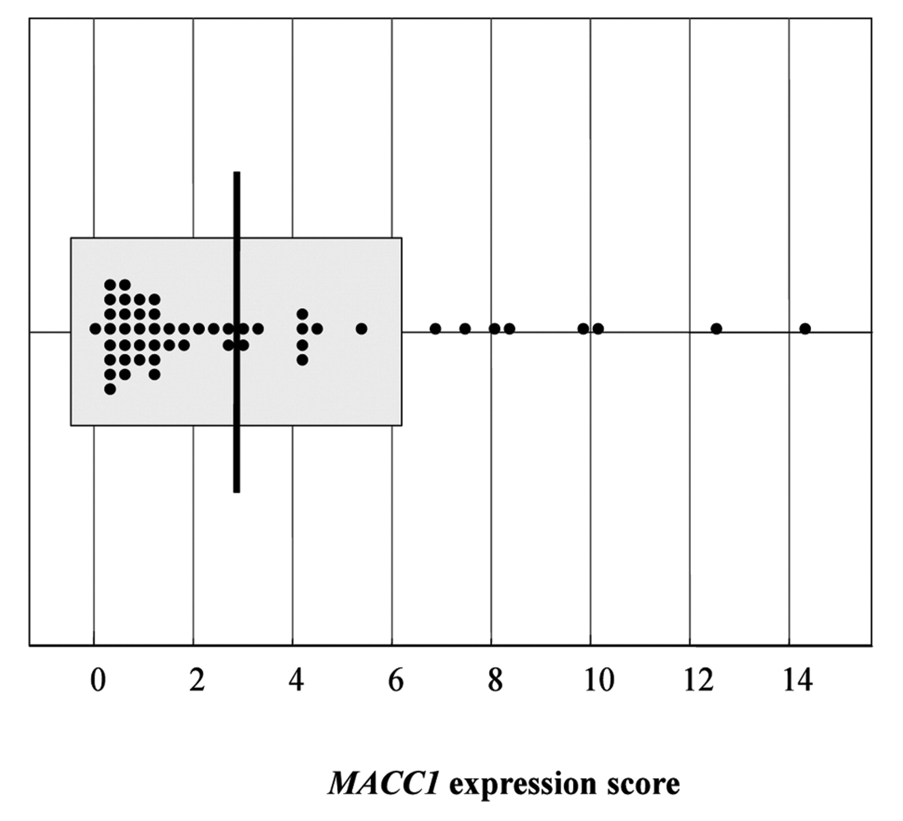
MACC1 expression scores were distributed between 0.11 and 14.26 (the average was 2.88±3.34).

MACC1 expression scores according to TNM stage. A significant increase in expression scores was observed in stage IV colorectal carcinomas (5.55±4.97) compared to stage I, II and III colorectal carcinomas (2.32±2.64) (p=0.007) .

MACC1 expression scores according to peritoneal dissemination. A significant increase in expression scores was observed in cases with peritoneal dissemination (5.75±4.58) compared to those without (2.57±3.09) (p=0.042).
We demonstrated that MACC1 expression was up-regulated according to the malignancy of colorectal cancer. Although the population study here was small, and further examination will be necessary, these results suggest that MACC1 might serve as a new parameter for the prognostic prediction of colorectal cancer.
Acknowledgements
We would like to thank M. Ogata for her technical assistance.
- Received January 26, 2010.
- Revision received May 26, 2010.
- Accepted June 3, 2010.
- Copyright© 2010 International Institute of Anticancer Research (Dr. John G. Delinassios), All rights reserved


