


Abstract
Background: Keratinocyte growth factor (KGF increases the proliferation and motility of many epithelial cells and is known to be up-regulated in pancreatic cancer. The present study examined the hypothesis that KGF may initiate or enhance the progression of pancreatic cancer by increasing the proliferation and motility of pancreatic cancer cells. Materials and Methods: HPAF-II pancreatic cancer cell migration and proliferation was evaluated using a culture wounding assay 24 and 48 hours following KGF treatment. KGF receptor (KGFR) localization in these cells was established by immunohistochemistry. Results: KGF treatment significantly increased the proliferation and motility of the HPAF-II cells. In addition, KGF enhanced the motile morphology of these cancer cells. Conclusion: The results of this study indicate that KGF has a rapid influence on the proliferation and motility of HPAF-II cells and suggest that KGF may be involved in the progression of pancreatic cancer.
Carcinoma of the pancreas is the fourth leading cause of cancer deaths in the United States. This malignancy is one of the most deadly, with a median survival of 6-10 months for patients with locally advanced disease and 3-6 months for those who have metastatic disease. Clearly there is an urgent need to better understand the etiology of this disease process and to identify and develop new therapeutic targets, therapeutic agents and biomarkers for the early diagnosis and treatment of this disease.
Keratinocyte growth factor (KGF) was discovered in 1987 as a soluble factor in stromal tissue that enhances the growth of epithelial cells (1). KGF, originally isolated from human embryonic fibroblasts, is a member of the fibroblast growth factor (FGF) family of polypeptide growth factors and has also been designated FGF-7 (2). Although not present in epithelial cells, KGF stimulates DNA synthesis, proliferation and migration of epithelial cells in pancreatic, breast, prostate and other tissue (3). These biological actions of KGF are thought to be involved in normal morphogenesis and tissue repair; however, there is evidence that overexpression of KGF and/or its receptor may be associated with cancer progression (4-6). Accordingly, overexpression of KGF, its receptor (KGFR), or both has been observed in colorectal, prostate and lung carcinomas (7-9).
We have reported that KGF produced a rapid scattering motility effect on human breast cancer cells (10, 11). The effects of KGF on the motile morphology of the cancer cells persisted for up to 48 hours following KGF treatment (11). Further, we observed that specific inhibition of KGF receptor (KGFR) expression and/or KGF activity eliminated the KGF-induced motility response (12). In the case of human pancreatic cancer, it has been demonstrated that KGF is overexpressed in pancreatic cancer tissue; furthermore, KGF enhances the proliferation of ductal epithelial cells (13, 14). This evidence appears to suggest that KGF, secreted from stromal tissue, may initiate or enhance the metastatic progression of pancreatic cancer. Thus, the goal of the present study was to examine the influence of KGF on the proliferation and motility of pancreatic cancer cells.
Materials and Methods
Cell culture methods. The human pancreatic cancer cell line (HPAF-II) used in this study was obtained from the ATCC and grown using our standard culture conditions (10, 11). This cell line has been characterized as a cell culture model for pancreatic cancer (15). Briefly, the cells were grown as monolayer cultures in RPMI-1640 media (without phenol red) supplemented with 2 mM L-glutamine, gentamicin (50 μg/ml), penicillin (100 units/ml), streptomycin (100 μg/ml), estradiol (10-11 M) (all from Sigma, St. Louis, MO, USA) and 5% bovine calf serum (Hyclone, Logan, UT, USA).
Cell migration assay. Cell migration and proliferation were evaluated using a culture wounding migration assay as previously described (11). Three days after seeding 5×10⁵ HPAF-II cells into 60 mm culture dishes, when the cells were approximately 80% confluent, the cells were wounded and treated with human recombinant KGF (R&D Systems, Minneapolis, MN, USA) at a concentration of 250 ng/ml. At 24 and 48 hours following treatment, cell migration was determined by measuring both the distance traveled by the cell front into the wounded area (migration index) and the number of cells in the wounded area (proliferation index)/microscopic field. Measurements were taken from 10-12 individual microscopic fields in each experiment and data were summarized from 2-3 experiments.

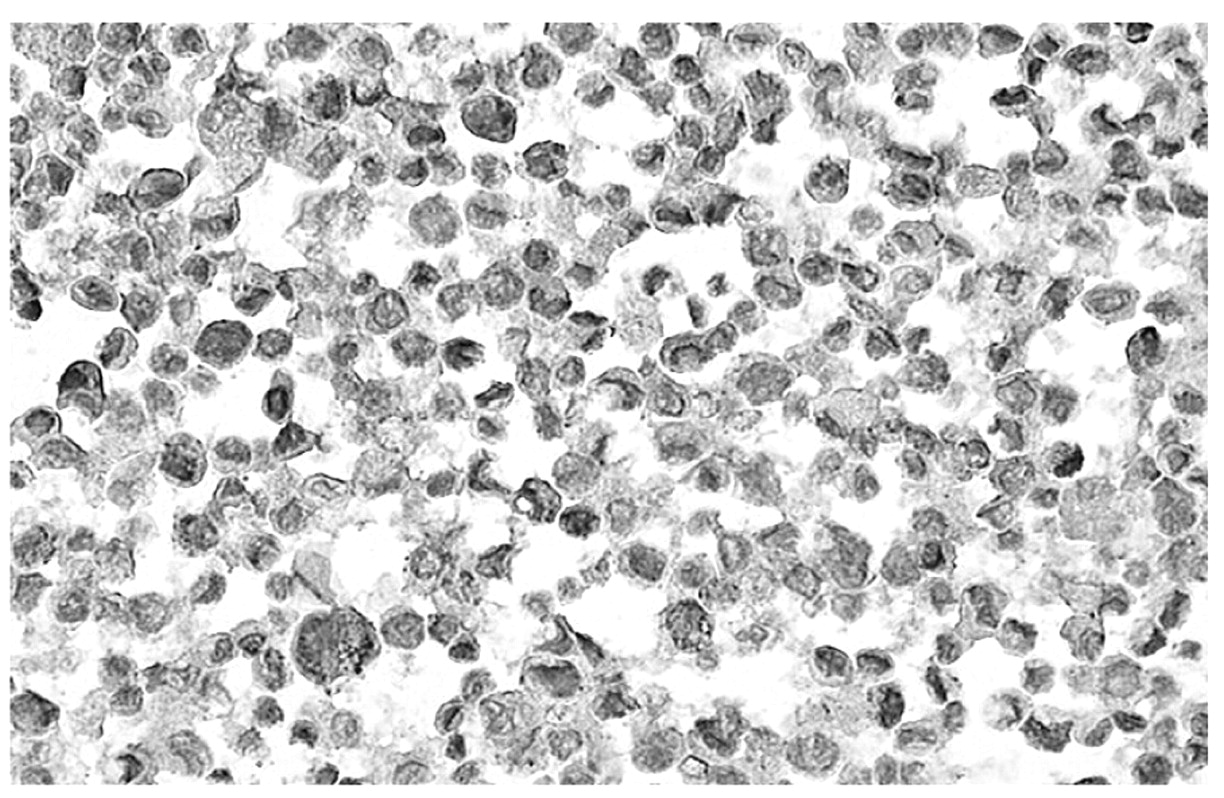
Immunolocalization of KGFR in HPAF-II cell culture. In this photomicrograph (×200), increased density represents KGFR localization. The cells were grown in control media.
Digital imaging. In this study, image analysis was conducted as previously described (11). Phase contrast images of cancer cells were recorded on an inverted microscope (×200-400) at the end of the 48-hour treatment period using a digital camera (Model DP70-BSW-V1.2; Olympus Corp., Center Valley, PA, USA) inserted into the optics.
Immunohistochemistry. Cells were fixed overnight in 0.1% formal saline and then pelleted and prepared for immuohistochemistry as previously described (16). Briefly, the pellet was resuspended in agarose, solidified and processed for paraffin embedding. Five-micron tissue sections were cut from paraffin-embedded blocks and processed for immunohistochemistry using the ImmPRESS Universal Reagent Kit (Vector Laboratories, Burlingame, CA, USA). Slides were rinsed extensively in PBS, treated with diluted normal blocking serum followed by a 2 hour incubation with KGFR Bek rabbit polyclonal antibody (1:50 dilution; Santa Cruz Biotechnology, Inc, CA, USA). Following incubation at room temperature, sections were washed in PBS, incubated in NovaRED™ (Vector Laboratories) and counterstained with ImmunoMaster Hematoxylin (AmericanMaster Tech Scientific, Inc., Lodi, CA, USA). Cells processed in the same manner, except without KGFR primary antibody, were included to exclude negative immunoreactivity.
Statistical analysis. Multiple group comparisons were conducted using ANOVA and Student's t-test for pair-wise comparisons. Group differences resulting in p-values of less than 0.05 were considered to be statistically significant.

Effect of KGF on HPAF-II cell proliferation and motility. A, The effect of KGF (250 ng/ml) on the number of HPAF-II cells migrating in wounded cultures. Each bar represents the mean cell number from 10-12 microscopic fields along the cell front ±SEM. B, Dose-response effect of KGF (250 ng/ml) on the distance traveled of migration of HPAF-II cells in wounded cultures. Each bar represents the mean distance of cell migration in 10-12 microscopic fields along the cell front ±SEM.
Results
In this study, the presence of KGFR in HPAF-II pancreatic cancer cells was confirmed by immunohistochemistry. These cancer cells stained positively for the KGFR as indicated by the density of hematoxylin stain (Figure 1). Furthermore, we observed that human recombinant KGF produced a significant increase in cell proliferation and motility at 24 and 48 hours after KGF administration in these pancreatic cancer cells (Figure 2A and B). In addition, KGF produced a change in the morphology of the wounded pancreatic cancer cell cultures which is consistent with that of highly invasive cancer cells (Figure 3A and B).
Discussion
HPAF-II is a metastatic pancreatic cancer cell line which is reported to be very invasive (17). Accordingly, this cell line has been employed by others to examine factors involved in pancreatic cancer progression and metastasis (18, 19). Thus, these cells were employed in the present study to examine the influence of KGF on the progression of pancreatic cancer.
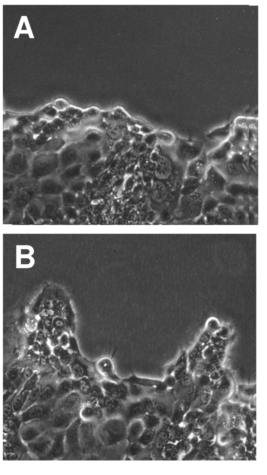
Effect of KGF on the morphology of HPAF-II cells in wounded cultures. Photomicrographs (×200) of wounded cell cultures at 48 h following treatment with control vehicle (A) or KGF (250 ng/ml) (B).
KGF expression and KGFR signaling appear to be associated with enhanced proliferation and the progression of human pancreatic cancer. For example, Siddiqi and coworkers observed that KGF expression was enhanced in 7 out of 16 human pancreatic cancer samples and that 5 out of 7 pancreatic cancer cell lines expressed KGFR (13). Similarly, overexpression of KGFR in 7 out of 10 pancreatic cancer samples was observed using a cDNA cancer profiling array comparing tumor and patient-matched normal tissue (20). Interestingly, it was reported that KGF and KGFR expression levels were increased 5-fold within 28 days in a rat model of pancreatitis in vivo (21). In addition, overexpression and co-localization of KGF and KGFR in pancreatic cancer and adjacent parenchyma has been observed indicating that KGF may enhance the progression of pancreatic cancer in either an autocrine or paracrine manner (22). Similarly, other FGFs and FGF receptors and are overexpressed and appear to be involved in the invasiveness of pancreatic cancer (23-25).
The present study demonstrated that the proliferation and motility of the HPAF-ll pancreatic cancer cell line was enhanced by KGF treatment within 24 to 48 hours. The results of the present and previous studies suggest that up-regulation of KGF secretion and/or KGFR expression may be early signals in the progression of the pancreatic cancer (13, 22, 26). Accordingly, KGFR expression levels may be a useful prognostic biomarker. Furthermore, if this hypothesis is correct, therapeutic inhibition of KGFR signaling and/or inhibition of genes and proteins regulated by KGF/KGFR signaling may impede the progression of pancreatic cancer cells to a more metastatic phenotype (25, 27).
Acknowledgements
This study was supported in part by grants from NIH/NCI (CA-125493) (CA-89740) and DoD (DAMD17-01-1-0591).
- Received February 25, 2009.
- Revision received April 27, 2009.
- Accepted May 5, 2009.
- Copyright© 2009 International Institute of Anticancer Research (Dr. John G. Delinassios), All rights reserved


