


Abstract
Background: Sonodynamic therapy (SDT) is a promising methodology for cancer treatment. Methylene blue (MB) is a phenothiazine dye that is widely used in clinical practice and can be administered intravenously. Materials and Methods: The sonodynamic antitumor effect of 1, 10 and 100 μM MB on sarcoma180 (S180) cells was investigated in vitro. Results: After ultrasound (US) exposure at 0.24 W/cm2 for 30 seconds, survival rates of S180 cells in the presence of 10 and 100 μM MB were significantly lower than that of the control group. These effects were significantly inhibited by the addition of D-mannitol, but not by L-histidine or superoxide dismutase. Microvilli loss and blebbing on the surface of S180 cells were observed in the presence of 100 μM MB after US exposure. Conclusion: MB has a sonodynamic antitumor effect on S180 cells in vitro and the hydroxyl radical appears to be the principal mediator of this effect.
Sonodynamic therapy (SDT) is a method by which cytotoxic effects of drugs (sonosensitizers) on tumor cells is enhanced by exposure to ultrasound (US) (1-5). Because US can be focused on a small region and can deeply penetrate tissue, SDT may prove to be a useful tool for the clinical treatment of tumors located deep within the body (6-11). Umemura and colleagues first described SDT in a study of the synergistic effect of US and hematoporphyrin (12, 13). In recent years, a number of studies on sonosensitizers have been reported (14-17). Hematoporphyrin and its derivatives exhibit a sonodynamic antitumor effect and accumulate in tumor tissues; however, these agents can cause photodermatitis. Sonosensitizers with low toxicity are desirable for clinical use.
Methylene blue (MB) is an inexpensive phenothiazine dye with low toxicity and has been approved for clinical use (Figure 1). MB has antifungal, antibacterial (18) and antimalarial activity (19). It also has been used to stain living organisms, to treat methemoglobinemia (20, 21), and to prevent ifosfamide-induced encephalopathy (22). Recently, MB was discovered to have photodynamic effects (23-26). In a previous study, it was demonstrated that acridine orange (AO) has a sonodynamic antitumor effect, in addition to its photodynamic properties (27). As MB has a very similar chemical structure to AO, it is believed that MB and AO may have similar physiological properties. However, there are no reports regarding the sonodynamic effect of MB. Therefore, the sonodynamic effect of MB on sarcoma 180 (S180) cells was investigated in vitro.
Materials and Methods
Preparation of tumor cells. Murine ascitic tumor cells (S180, Medical Cell Resource Center, Tohoku University Gerontology Research Institute, Sendai, Japan) were used as the experimental tumor. A suspension of approximately 1 mL S180 cells was injected intraperitoneally into 7-week-old ICR male mice (Japan SLC, Inc., Shizuoka, Japan). Approximately 7 to 14 days later, 1 to 2 mL of ascitic fluid was collected and diluted in phosphate-buffered saline (PBS) to a cell density of 1.0×10⁶/mL. The survival rate of tumor cells was evaluated by the trypan blue dye exclusion method using a hemocytometer (Kayagaki, Tokyo, Japan) under an optical microscope (Olympus BH-210, Tokyo, Japan; ×400). Viability before treatment was always greater than 98%.
Drug. MB was purchased from Nacalai Tesque Inc. (Kyoto, Japan). The MB-containing solutions were prepared by dissolving MB in PBS to provide 2, 20 and 200 μM solutions.
Ultrasound apparatus. The US apparatus used in this study was manufactured at the Department of Electronic Engineering, Akita University Mining College, Akita, Japan. The generator consists of a ceramic, cylindrical transducer combined with a function generator. It can be used at a resonance frequency of 2 MHz on a piezo-electric element. A round, ceramic plate measuring 20 mm in diameter and 1 mm in thickness (2Z 20D SYIC; Fuji Ceramics, Shizuoka, Japan) was used as the piezo-electric element. The resonance frequency after adhesion to aluminum was 2.26 MHz. A function generator (FG-350; Iwatsu Denshi, Tokyo, Japan) capable of operation over a frequency bandwidth of 0.1 Hz to 10 MHz was used. The sine-wave mode was used in this experiment. The system also included a power amplifier (PA 40-2801; Someway, Shizuoka, Japan) with a frequency bandwidth of 100 kHz to 350 MHz and an output of 0 to 10 W, and a power meter (SX-200; Daiichi Denpa Kogyo, Tokyo, Japan) with a frequency range of 1.8 to 200 MHz and a power measurement range of 0 to 200 W (Figure 2).
Preparation of the experimental tumor fluid. For each experiment, the tumor fluid was prepared by mixing 0.7 mL of cell suspension with either 0.7 mL of MB-containing solution (2, 20 or 200 μM) or 0.7 mL of PBS as control. The final concentrations of MB-containing solutions were 1, 10 and 100 μM. These solutions were introduced separately into a glass cell that was 20 mm in diameter, 25 mm in height, and had a base of 9-μm-thick Mylar film (glass cell, manufactured at the Instrument Center, Akita University School of Medicine, Akita, Japan; Mylar film, Teijin DuPont Films Co., Tokyo, Japan). The density of tumor cells contained in each glass cell was set at approximately 1.0×10⁶/mL, as indicated earlier. Initially, the survival rate of tumor cells in the presence of 100 μM MB, without US exposure (n=8), was examined.
Ultrasound exposure experiment. The control (PBS) and 1, 10 and 100 μM MB-containing solutions were exposed to US at 0.24 W/cm2 at a frequency of 2 MHz for 15, 30 or 60 seconds (n=8). In addition, the control and 1, 10 and 100 μM MB-containing solutions were exposed to US at 0.18, 0.24 or 0.36 W/cm2, 2 MHz, for 30 seconds (n=8). To ensure close adhesion of the piezo-electric element with the glass cell, transmission gel (US FINE GEL; Fukuda Denshi, Tokyo) was applied. All procedures in the US exposure experiment were performed within 1 hour after aspiration of ascitic fluid from mice. The temperature of the solution in the glass was set at room temperature (22-26°C). During the sonication procedure, the temperature inside the glass cell did not rise by more than 2°C, as measured by a digital thermometer (TESTO 905-T1, Kanagawa, Japan). Cell survival rate in the experiment was defined as number of living cells after US exposure/number of living cells before US exposure ×100 (%); cells that were destroyed by US exposure were counted as dead cells.
Identification of active oxygen. L-histidine, D-mannitol and superoxide dismutase (SOD) were purchased from Wako Chemical Company (Tokyo, Japan). The survival rate of tumor cells was determined after US exposure at 0.24 W/cm2, 2 MHz, for 30 seconds (n=8) in the presence of 100 μM MB, with or without active oxygen scavengers. One hundred μg/mL SOD was used as a H2O2 scavenger, 50 mM L-histidine hydrochloride monohydrate was used as a singlet oxygen and hydroxyl radical scavenger, and 100 mM D-mannitol was used as a hydroxyl radical scavenger.
Electron microscopy. Before and immediately after US exposure (0.24 W/cm2, 2 MHz, 30 seconds) in the presence or absence 100 μM MB, the cells were fixed in 3.0% glutaraldehyde solution buffered to pH 7.2. The cells were then gradually dehydrated with increasing concentrations of ethanol before being transferred to a freeze-drier, coated with gold and examined with a 1200 EX scanning electron microscope (Electron-microscope Optical Ltd. JOEL, Japan) at 10 kV.

Chemical structure of methylene blue (MB).
Statistical analysis. The mean and standard deviation of the survival rate of tumor cells was calculated for each group. Differences between groups were considered significant when the p-value on Dunnet's test was 0.05 or lower.
Results
Cell damage. Without US exposure the survival rate of S180 cells in the presence of 100 μM MB was not significantly different from that of the control group within 1 hour, and there were no significant differences in survival rate among MB-containing groups (data not shown). The survival rates of S180 cells in both the control and the MB-containing groups at an intensity of 0.24 W/cm2 declined as US exposure time increased. At a US exposure time of 15 seconds, the survival rate in the presence of 100 μM MB was significantly lower than that of the control group. At 30 seconds, the survival rates in the presence of 10 and 100 μM MB were significantly lower than that of the control group. At 60 seconds, the survival rate in the presence of 100 μM MB was significantly lower than that of the control group. However, the survival rate was very low even in the control group at 60 seconds (Figure 3).
The survival rate of S180 cells in both the control and the MB-containing groups at a US exposure time of 30 seconds declined as US intensity increased. At an intensity of 0.18 W/cm2, survival rates were not significantly different among the control group and MB-containing groups. At 0.24 W/cm2, the survival rates in the presence of 10 and 100 μM MB were significantly lower than that of the control group. At 0.36 W/cm2, the survival rates in both the control and MB-containing groups were very low and no significant differences in survival rates were observed between the control and the MB-containing groups (Figure 4).
Identification of active oxygen. The sonodynamic effect of 100 μM MB was significantly lower in the presence of D-mannitol, as compared with MB alone, but not in the presence of L-histidine or SOD (Figure 5).
Scanning electron microscope observations. In the control group without US exposure, cells were round or oval with numerous microvilli over the surface of the cells (Figure 6A). In the 100 μM MB-containing group without US exposure, no obvious cell damage was observed (Figure 6B). Cells in the control group exposed to US were mostly spherical in shape; however, the number of microvilli was lower, and some blebs had formed on the cell surface (Figure 6C). Cells exposed to US in the presence of 100 μM MB were irregularly shaped; most microvilli had disappeared, and numerous blebs of various sizes had formed on the cell surface (Figure 6D).
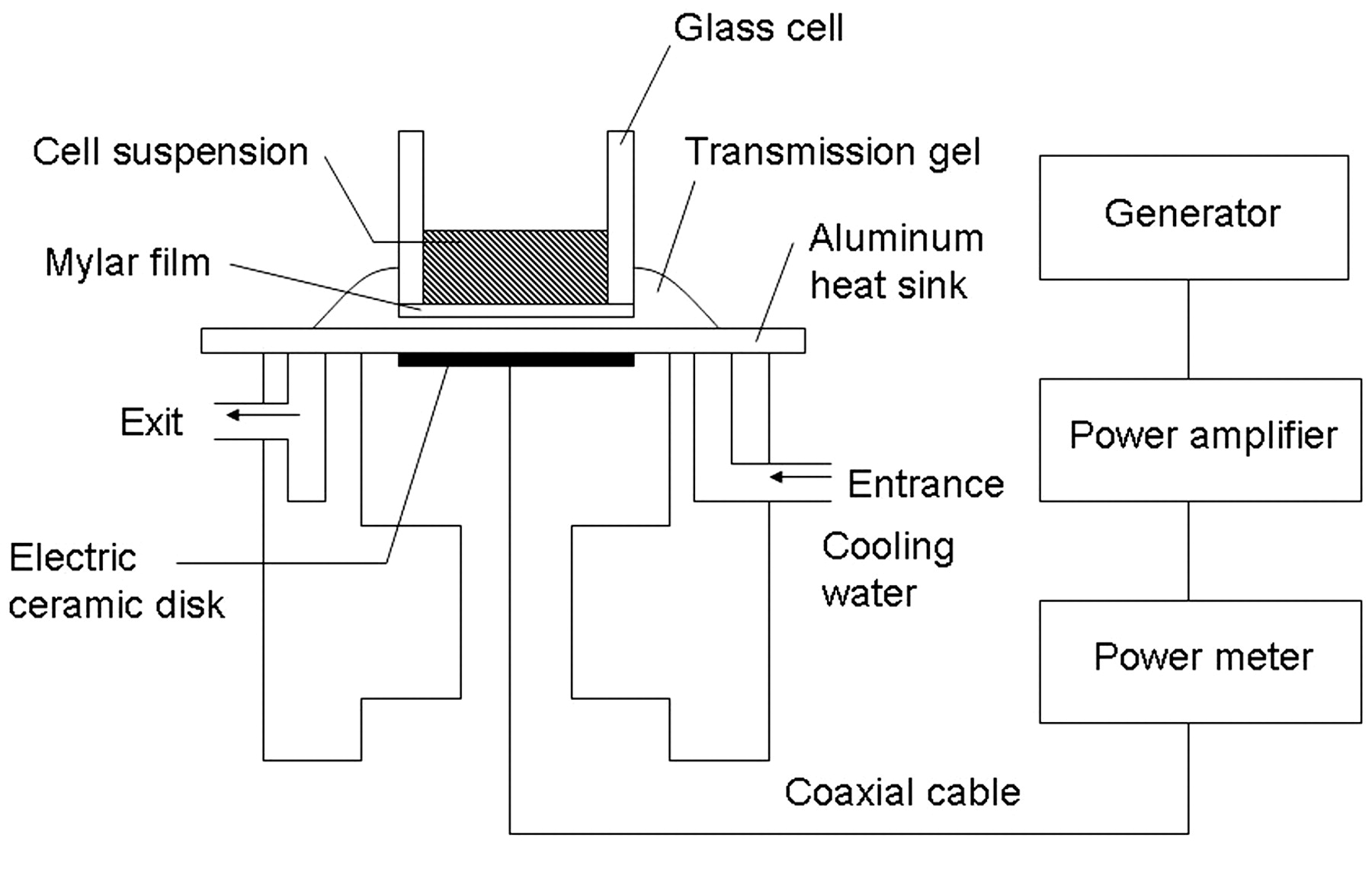
Overview of the US apparatus.
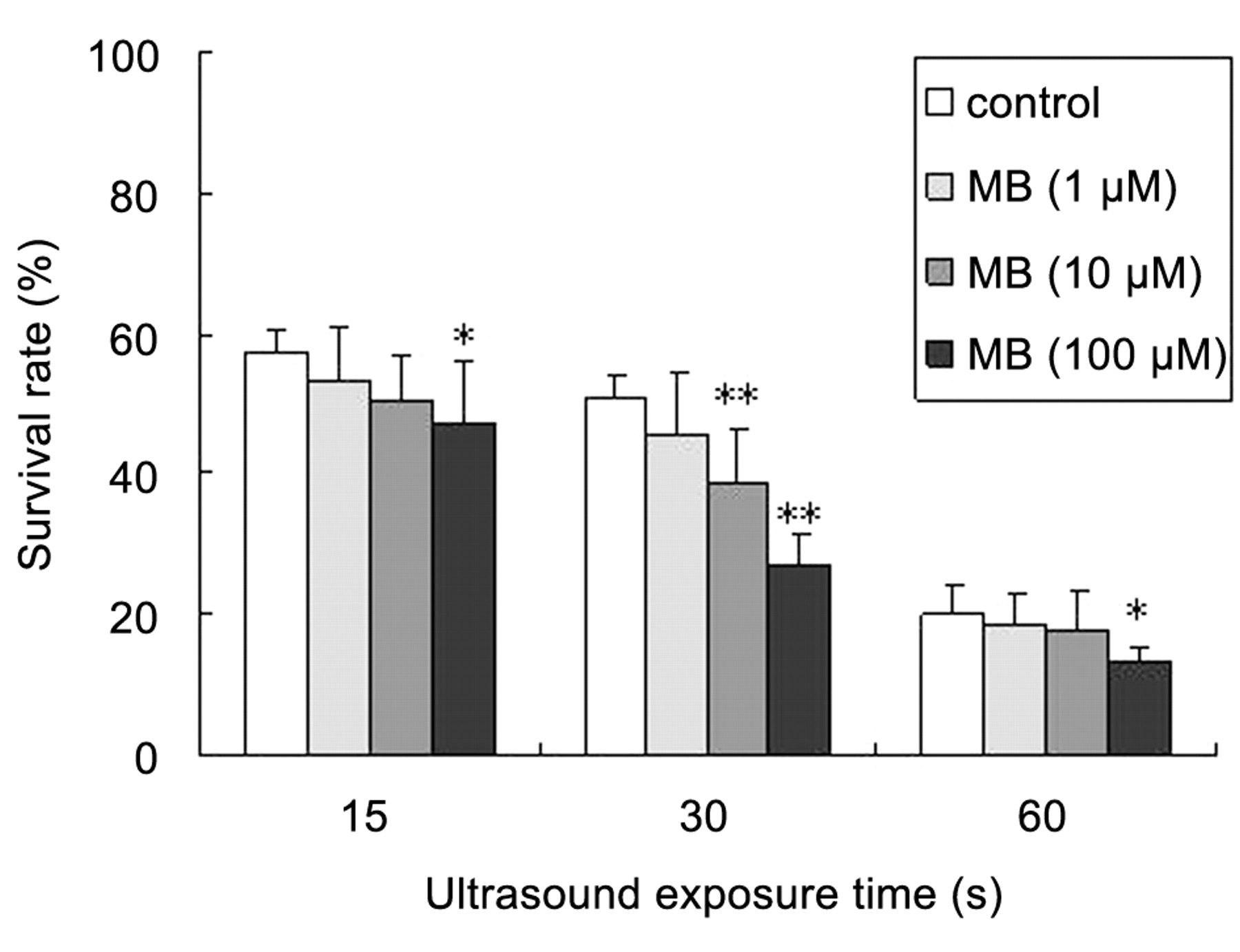
Effect of US exposure on S180 cells for various exposure times at an intensity of 0.24 W/cm2 (mean±SD, n=8). *p<0.05; **p<0.01 vs. control group.
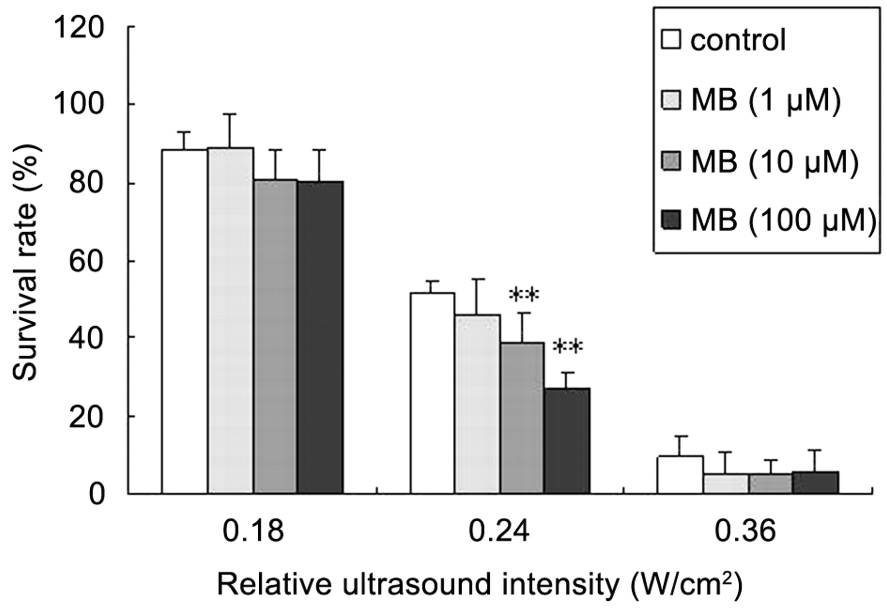
Effect of US exposure on S180 cells at various US intensities for an exposure time of 30 seconds (mean±SD, n=8). **p<0.01 vs. control group.
Discussion
In this study, it is demonstrated that MB exerted a dose-dependent sonodynamic effect on S180 cells in vitro. The survival rate in the presence of 100 μM MB was significantly lower than that of the control group at a US exposure time of 15 seconds; in the presence of 10 μM MB, the same result was obtained at 30 seconds (Figure 3). At 0.24 W/cm2, the survival rates in the presence of 10 and 100 μM MB were significantly lower than that of the control group (Figure 4). The sonodynamic antitumor effect on S180 cells was observed in the control group at 0.24 W/cm2 for 15-60 seconds. However, the effect was enhanced in the presence of MB. This may be due to the synergistic effect of US and MB. In a previous study, it was reported that sparfloxacin at a concentration greater than 200 μM had a sonodynamic effect at 0.24 W/cm2 for 30 seconds, and that 3.3 μM AO had an effect at 0.24 W/cm2 for 60 seconds. The sonodynamic effect of MB is similar to that of AO and stronger than that of sparfloxacin. These findings indicate that MB has potential for clinical use in SDT.

Effect of active oxygen scavengers on cell damage in the presence of 100 μM MB after US exposure at an intensity of 0.24 W/cm2 for 30 seconds (mean±SD, n=8). *p<0.01 vs. no-scavenger group.
The sonodynamic effect of MB was significantly reduced by D-mannitol; although no obvious change was observed in the presence of either L-histidine or SOD (Figure 5). These findings suggest that the hydroxyl radical is an important mediator of the sonodynamic antitumor effect of MB. The sonodynamic effect of sparfloxacin was reduced by L-histidine, and the effect of AO was reduced by L-histidine and D-mannitol (3, 27). In the context of these previous results, the role played by singlet oxygen in cell damage in SDT using MB remains obscure.

Scanning electron microscopic images of S180 cells. A, Cells in the control group without US exposure; B, cells in the 100 μM MB-containing group without US exposure; C, cells in the control group after US exposure at 0.24 W/cm2 for 30 seconds; D, cells in the 100 μM MB-containing group after US exposure at 0.24 W/cm2 for 30 seconds.
In scanning electron microscope observations, ultrasonically induced cell membrane damage was much more serious in the presence of 100 μM MB than in the control group, which is consistent with the findings observed with sparfloxacin (4). Changes on the surface of the cell membrane affect membrane function and eventually lead to cell death.
Many drugs have been investigated as possible sono-sensitizers, but none are approved for clinical use in SDT. MB is widely used in clinical practice and can be administered by intravenous injection. Matsubara et al. (23) described the photodynamic effects of MB for osteosarcoma in vitro and in vivo studies. They noted that MB in photodynamic therapy had a strong cytocidal effect on mouse osteosarcoma cells in vitro, but did not inhibit tumor growth in vivo. Tumor selectivity and the sonodynamic effects of MB in the treatment of tumor-bearing mice should be further investigated.
In conclusion, because of its excellent sonodynamic effect and low toxicity, MB has promise as a sonosensitizer for SDT.
- Received December 2, 2008.
- Revision received January 17, 2009.
- Accepted March 11, 2009.
- Copyright© 2009 International Institute of Anticancer Research (Dr. John G. Delinassios), All rights reserved


