


Abstract
An increased incidence of colorectal carcinoma is known to occur in patients with ulcerative colitis, which displays a cycle of recurrence-remission in the colorectal mucosa. Repeated oral doses of 3% dextran sulfate sodium subsequent to a single intraperitoneal injection of azoxymethane induces chronic ulcerative colitis, resulting in an increased incidence of high-grade dysplasia and submucosal-invasive adenocarcinomas in the mouse colorectum. The features of the colitis induced in this animal model are very similar to the ulcerative colitis in patients in terms of both clinical and histopathological characteristics. Bisphosphonates are known to increase bone mass by suppressing bone turnover in postmenopausal women. A novel single-nitrogen bisphosphonate, ibandronate, is effective in preventing skeletal events in patients with bone metastases from colorectal cancer. Decreasing the bone mineral affinity of bisphosphonates is an effective therapeutic strategy to inhibit skeletal tumor growth in vivo. In the present study, the preventative effects of the bisphosphonate ibadronate on colorectal carcinogenesis in mice treated with azoxymethane and dextran sulfate sodium was investigated. Additive treatment with bisphosphonate prevented the shrinkage of colorectum which was affected by a cycle of recurrence-remission in colorectal mucosa, resulting in a reduced incidence of colorectal dysplasia and a reduced expression of thymidine kinase mRNA in the colorectum. Taken together, the present results indicate that ibadronate may inhibit colorectal carcinogenesis and its development by inhibiting colorectal epithelial cell proliferation and the neoplastic process.
Bisphosphonate (BP) is known to increase bone mass by suppressing bone turnover in postmenopausal women (1, 2). One BP, ibadronate, sodium cycloheptylaminomethylene-bisphosphonate monohydrate, is also known to be an antiresorptive agent without inhibiting bone formation (3, 4). As previously reported (5), the chemical castration of rats using the gonadotropin-releasing hormone (GnRH) agonist leuprorelin acetate for 16 weeks reduced the bone mineral content (BMC) value to 91.0% of that of the normal control animals. However, a simultaneous 8-week administration of the BP markedly enhanced the BMC value to 110.1% of that in the rats treated with the GnRH agonist alone. It was reported that the addition of zoledronic acid, a third-generation BP, to adjuvant endocrine therapy improved disease-free survival in premenopausal patients with estrogen-responsive early breast cancer (6). Zoledronic acid showed potent antitumoral and apoptosis-inducing effects on HCT-116 colon cancer cells (7).
An increased incidence of colorectal carcinoma is known to occur in patients with ulcerative colitis (UC) (8), which displays a cycle of recurrence-remission, i.e. periods of ulceration and regeneration of the colorectal mucosa. As previously reported (9), three administrations of 3% dextran sulfate sodium (DSS) subsequent to a single intraperitoneal injection of azoxymethane (AZM) induced chronic UC, resulting in an increase of high-grade dysplasia and submucosal-invasive adenocarcinomas in mouse colorectum. The features of the colitis induced in this animal model are very similar to those in patients in terms of both clinical and histopathological characteristics, i.e. diarrhea, occult blood, melena, mucosal inflammatory cell infiltration, crypt abscess formation and mucosal erosion (10).
Thymidine kinase (Tk2; EC 2.7.1.21) and thymidylate synthase (Tyms; EC 2.1.1.45) catalyze the formation of deoxythymidine monophosphate (dTMP) by the phosphorylation of thymidine via the salvage pathway, and by the methylation of deoxyuridine monophosphate (dUMP) with the concomitant conversion of N5,N10-methylenetetra-hydrofolic acid to 7,8-dihydrofolic acid via the de novo pathway, respectively (11). High Tk2 and Tyms activities have been found in rapidly proliferating tissues of normal, fetal and neoplastic tissues (12-14). In the present study, the preventative effects of BP on colorectal carcinogenesis in mice treated with AZM and DSS was investigated.
Materials and Methods
Animals and chemicals. Forty-five specific pathogen-free female CBA/J mice (Charles River Japan, Tokyo, Japan), 6 weeks of age, were used. The animals were housed in plastic cages with steel shavings under controlled temperature (24±0.5°C) and lighting (12 h of light from 0600 to 1800 h), and were permitted free access to a commercial diet (CE-2, CLEA Japan, Tokyo, Japan) and tap water at the animal research center of Tokyo Medical and Dental University (Tokyo, Japan).
At 8 weeks of age, the animals were divided into 3 groups of 15 mice each: one control group (Normal-Control) and two experimental groups (AZM/DSS-Control and AZM/DSS-BP). The animals of the two experimental groups (30 mice) were injected intraperitoneally with 8.0 mg/kg AZM (Sigma Chemical, St Louis, MO, USA). In the control group, 15 mice received 0.1 ml of a 0.9% NaCl solution by the same procedure. Two weeks after the intraperitoneal pretreatment with AZM, the animals in the two experimental groups were given distilled water containing 3% (w/v) synthetic DSS (Mol. Wt. 50,000; Ensuiko Sugar Refining, Yokohama, Japan) for 7 days followed by tap water for 14 days, a total of three times. Beginning at 8 weeks of age, the animals in one of the two experimental groups were fed the same commercial diet containing ibadronate (250 mg/kg diet: YM175; a gift from Yamanouchi Pharmaceuticals, Tokyo, Japan) as a BP for 12 weeks (AZM/DSS-BP group). The other 30 mice in the Normal-Control and AZM/DSS-Control groups received the same commercial diet alone for 12 weeks.
Experimental procedures and measurements. Changes in body weight were recorded every week throughout the experiment. All animals were anesthesized with ether, and sacrificed by cervical dislocation at 20 weeks of age, and the liver, spleen, kidney, uterus, adrenals, ovaries and colorectum were removed and weighed. All experimental procedures conformed to the regulations described in the U.S. National Institutes of Health (NIH) Guide to the Care and Use of Laboratory Animals.
The longitudinal length of each colorectum was measured, and each specimen was longitudinally sectioned into two parts: one was stored at −80°C until evaluation of the expression levels of Tyms and Tk2 mRNA at −80°C and the other was immediately fixed in a 10% formaldehyde buffer solution (pH 7.2), embedded in paraffin, and prepared as 5 μm serial sections, then stained with Mayer's hematoxylin and eosin for histological examination.
Analysis of gene expression levels of Tyms and Tk2 in the colorectum. RT-PCR was performed for quantitative analysis of Tyms and Tk2 mRNA levels in the colorectum. Total RNA was extracted from each colorectal sample with a QuickPrep™ Total RNA Extraction Kit (Amersham Pharmacia Biotech, Buckinghamshire, UK). Reverse transcription was performed using oligo (dT) primers [0.5 ml oligo (dT)12-18 primers (1.0 mg/ml) (GIBCO BRL, Gaithersburg, MD, USA)] with a SUPERSCRIPT™ Preamplification System (GIBCO BRL) according to the supplier's instructions. Once the cDNA copy had been created using the mRNA template, the PCR was conducted immediately, as outlined below. Alternatively, the cDNA was stored at −20°C until use. The PCR was performed with recombinant Taq DNA polymerase (Nippon Gene, Tokyo) according to the manufacturer's instructions. The RNA (1.0 mg) was subjected to RT-PCR using the primers for Tyms and Tk2 cDNA for 34 cycles (each cycle consisted of denaturing at 94°C for 40 seconds, annealing at 55°C for 40 seconds and extension at 72°C for 40 seconds) in a Gene Amp PCR System 2400 (Perkin Elmer, Branchburg, NJ, USA). RT-PCR was carried out with three sets of primers (Tyms: 5′-TGAATGGGGAGCTATCTTGCCA-3′ and 5′-TCGTTG-GATGTGG ATTATACCC-3′; Tk2: 5′-TAGCACAGGCGGCACACGGAGT-3′ and 5′-TGCTCCGCGATGTGACCCAGGA-3′; and β-actin: 5′-AGGCCCAGAGCAAGAGAG-GCAT-3′ and 5′-CATGGCTGGGG TGTTGAAGGTC-3′). The levels of Tyms mRNA and Tk2 mRNA were determined by densitometry from photographs taken with an image analyzer (AE6920-MF Densitograph, ATTO, Tokyo), and are expressed as a ratio of the mRNA level of β-actin as an internal standard −80°C.
Calculations and statistics. All parameters were expressed as the mean±SEM. Statistical analysis was performed using the unpaired t-test, and Fisher's exact probability test. A p-value less than 0.05 was considered statistically significant.
Results
AZM/DSS treatment lowered the final body weight to 88.9% of that of the Normal-Control group (p<0.05) (Table I). The additive treatment with BP improved body growth despite AZM-DSS treatment, though not significantly. AZM/DSS treatment markedly altered organ weights, i.e. the weights of liver (p<0.05), spleen (p<0.01), kidney (p<0.05) and adrenals (p<0.01) were augmented compared with those of the Normal-Control group, although the weights of uterus and ovaries were reduced (Table I). However, the additive treatment with BP lowered the weights of spleen (p<0.01) and kidney (p<0.05), and elevated the weights of ovaries (p<0.01). The colorectal length in AZM/DSS-treated mice (AZM/DSS-Control group) was markedly reduced to 80.2% of that in the Normal-Control group (p<0.01) (Table I). The additive treatment with BP prevented the shrinking of the colorectum (p<0.01).
No open ulcer was found since mice in the experimental groups were sacrificed 3 weeks after the last administration of DSS. However, the number of foci of gland loss with inflammatory cell infiltration, i.e. indicating the severity of UC, in mice treated with BP (AZM/DSS-BP group) was reduced compared with that in the AZM/DSS-treated mice. AZM/DSS treatment induced high-grade dysplasia (Figure 1B), i.e. 27.9 sites/mouse in number and 184.1 mm2/mouse in cumulative area, although high-grade dysplasia was not found in the Normal-Control group (Figure 1A). However, the additive treatment with BP markedly reduced the high-grade dysplasia in both number and area to 7.2% and 4.5% of those in the AZM/DSS-Control group, respectively (p<0.01) (Figure 2). In the present study, no submucosal-invasive adenocarcinomas were found in any colorectal sample.
Growth, organ weights and colorectal length in mice of the study groups.
Expression levels of Tyms and Tk2 mRNAs in the whole colorectum in the AZM/DSS-Control group were markedly elevated to 2.1- and 9.8-fold the levels in the Normal-Control group, respectively (p<0.01) (Figure 3). However, the additive treatment with BP significantly lowered the gene expression level of Tk2, but not Tyms, to less than 60% of that in the AZM/DSS-Control group (p<0.05).
Discussion
AZM is known as a procarcinogen, which becomes an alkylating agent with carcinogenic activity following metabolic activation in the host (15). DSS is a synthetic, sulfated polysaccharide that induces colitis in rodents, which clinically and histologically resembles that in human UC. The hyperproliferation of cells in the inflammation-associated damage-regeneration cycle has been shown to contribute to the fixation of genetic and epigenetic alterations and promote the development of colorectal dysplasia and carcinoma (16). DSS tested negative in the Ames test for mutagens (17). However, nine cyclic administrations of DSS induced 9 low- and 4 high-grade dysplasias, and 2 carcinomas in 25 mice in our previous study (18). Inflammation-associated regenerative atypia is thought to be difficult to differentiate from dysplasia. Our histological diagnosis was supported by the findings of diffuse labelling of tumor cells with bromodeoxyuridine (BrdU) and activities of Tyms and Tk2 throughout the colorectal mucosa, i.e. BrdU uptake and activities of both Tyms and Tk2 in mucosal tumors were higher than in non-tumorous tissues (9). Thus, structural and cellular atypia pointed to a diagnosis of high-grade dysplasia. Accelerated epithelial cell turnover caused by chronic inflammation and epithelial damage might predispose the mucosa to DNA damage. AZM/DSS reduced body growth to 90% of the control, but the additive treatment with BP slightly increased growth despite the AZM/DSS treatment. Although AZM/DSS treatment increased the weight of organs except the ovary and uterus, BP had a tendency to normalize the altered weights except for the adrenals. Additive treatment with BP prevented the shrinkage of colorectum which was affected by a cycle of recurrence-remission in colorectal mucosa, resulting in a reduced incidence of colorectal dysplasia and a reduced expression of Tk2 mRNA in the colorectum.

Histopathological structure of colorectal tumorous region with high-grade dysplasia (B) (original magnification ×200) in a mouse treated with azoxymethane and 3% dextran sulfate sodium compared with normal mucosa (A) (×400) (H&E).
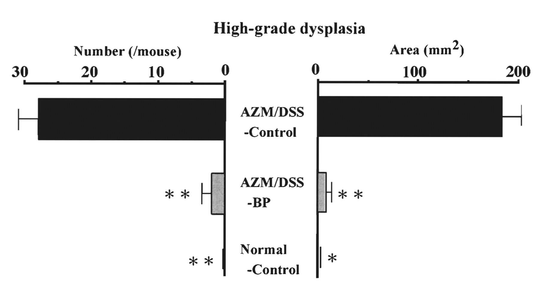
High-grade dysplasia in the colorectum: number (left) and area (right) in each group. Data are means±SEM. Significantly different from AZM/DSS-control at *p<0.05 and **p<0.01. AZM, Azoxymethane; DSS, dextran sulfate sodium; BP, bisphosphonate.

Expression levels of thymidylate synthase (Tyms) mRNA (left) and thymidine kinase (Tk2) mRNA (right) as a ratio of the β-actin mRNA level in the colorectum. Data are means±SEM. Significantly different from AZM/DSS-control at **p<0.01. AZM, Azoxymethane; DSS, dextran sulfate sodium; BP, bisphosphonate.
Heras et al. reported that a novel single-nitrogen BP, ibandronate, was effective in preventing skeletal events in patients with bone metastases from colorectal cancer (19). Fournier et al. demonstrated that decreasing the bone mineral affinity of BPs was an effective therapeutic strategy to inhibit skeletal tumor growth in vivo (20). Roelofs et al. have mentioned that nitrogen-containing BPs act intracellularly by inhibiting farnesyl diphosphate synthase, an enzyme of the mevalonate pathway, thereby preventing prenylation of small GTPase signaling proteins required for normal cellular function, and inhibition of farnesyl diphosphate synthase also seems to account for their antitumor effects observed in vitro and for the activation of γ,δ T-cells, a feature of the acute-phase response to BP treatment in humans (21). Taken together, our and other studies may indicate that BPs inhibit colorectal carcinogenesis and its development as an inhibitor of colorectal epithelial cell proliferation and the neoplastic process, or as a promoter of cellular differentiation.
Acknowledgements
The authors would like to express their cordial thanks to Professor Dr. Isao Okayasu from the Department of Pathology, School of Medicine, Kitasato University, Kanagawa 228-8555, Japan.
Footnotes
-
Competing Interests
The authors declare that they have no competing interests.
-
Funding
The present study was supported by the Foundations from Koihei Co. Ltd., Saitama, Japan and Japan Royal Jelly Co. Ltd,, Tokyo, Japan. The authors declare that there is no conflict of interest that would prejudice the impartiality of this scientific work.
- Received June 18, 2009.
- Revision received September 24, 2009.
- Accepted September 28, 2009.
- Copyright© 2009 International Institute of Anticancer Research (Dr. John G. Delinassios), All rights reserved


