


Abstract
Vitamin D has anti-proliferative and pro-apoptotic effects on several cell types, including breast cancer cells. There have been no studies quantifying the expression of the enzyme 25-hydroxyvitamin D-1-α-hydroxylase (1αOHase), which converts 25-hydroxyvitamin D to its active metabolite, in breast tissue. We performed real-time RT-PCR to quantity 1αOHase and vitamin D receptor (VDR) mRNA in samples of breast cancer, adjacent non-cancerous tissue and normal breast tissue. 1αOHase and VDR mRNA were universally expressed, however, 1αOHase expression was significantly down-regulated in adjacent non-cancerous tissue from women with breast cancer in comparison to individuals without cancer. VDR was also up-regulated in breast tumours. The vitamin D axis expression in the breast suggests a role for its actions in normal tissue homeostasis and breast cancer pathogenesis. The decreased expression of 1αOHase in normal tissue from women with breast cancer may be important in their predisposition to the development of cancer.
Epidemiological, animal and in vitro studies have demonstrated that vitamin D plays an important role in breast tumorigenesis (1). The main ligand for the vitamin D receptor (VDR) is 1,25(OH)2D, which is converted from its precursor 25(OH)D by 25-hydroxyvitamin D-1-α hydroxylase (1αOHase). Binding of 1,25(OH)2D to the VDR results in its translocation to the nucleus where it acts as a transcription factor. Extra-renal sites of 1αOHase expression have been described but its in vivo expression in the human breast has not been investigated. We have previously shown that 1αOHase mRNA levels are significantly lower in normal colonic tissue adjacent to the colon cancer compared to normal tissue from non-cancer patients (2), suggesting a possible genetic predisposition to colon cancer. Our aim was to determine whether a similar pattern in the breast might could provide a molecular explanation for the observed effects of vitamin D.
Materials and Methods
Thirty paired samples of tumour (T) and adjacent normal breast tissue (AN) and 18 samples of normal breast tissue from healthy patients undergoing benign breast surgery (NN) were studied. Local Ethical Committee approval was obtained from the East London and City Health Authority and all patients gave informed consent. RNA was isolated, quantified and its quality assessed as previously described (3). Primers and probes were designed using Primer Express (Applied Biosystems, Warrington, UK) and synthesized by MWG Biotech (Ebersberg, Germany). Their sequences are shown in the legend to Figure 1. mRNA levels were quantified on a Stratagene Mx4000 instrument using real time RT-PCR assays based on 5′ nuclease chemistry. Copy numbers were calculated relative to amplicon-specific standard curves obtained by serial dilution of single stranded sense oligonucleotides and reported as copy numbers/μg total RNA. Immunohistochemistry for the VDR was performed on 3 μm sections of paraffin-embedded blocks of the same samples using a standard avidin biotin complex method (Vector Laboratories) and 3′3′-diaminobenzidine tetrahydrochloride (DAB). Sections were incubated with a rabbit anti-human monoclonal VDR antibody at a 1/200 dilution. Specificity was confirmed by complete absence of staining by pre-incubation of the antibody with VDR ligand. Sections of kidney were used as a positive control in each run. The sections of normal breast and cancer were randomly mixed prior to staining.
Results
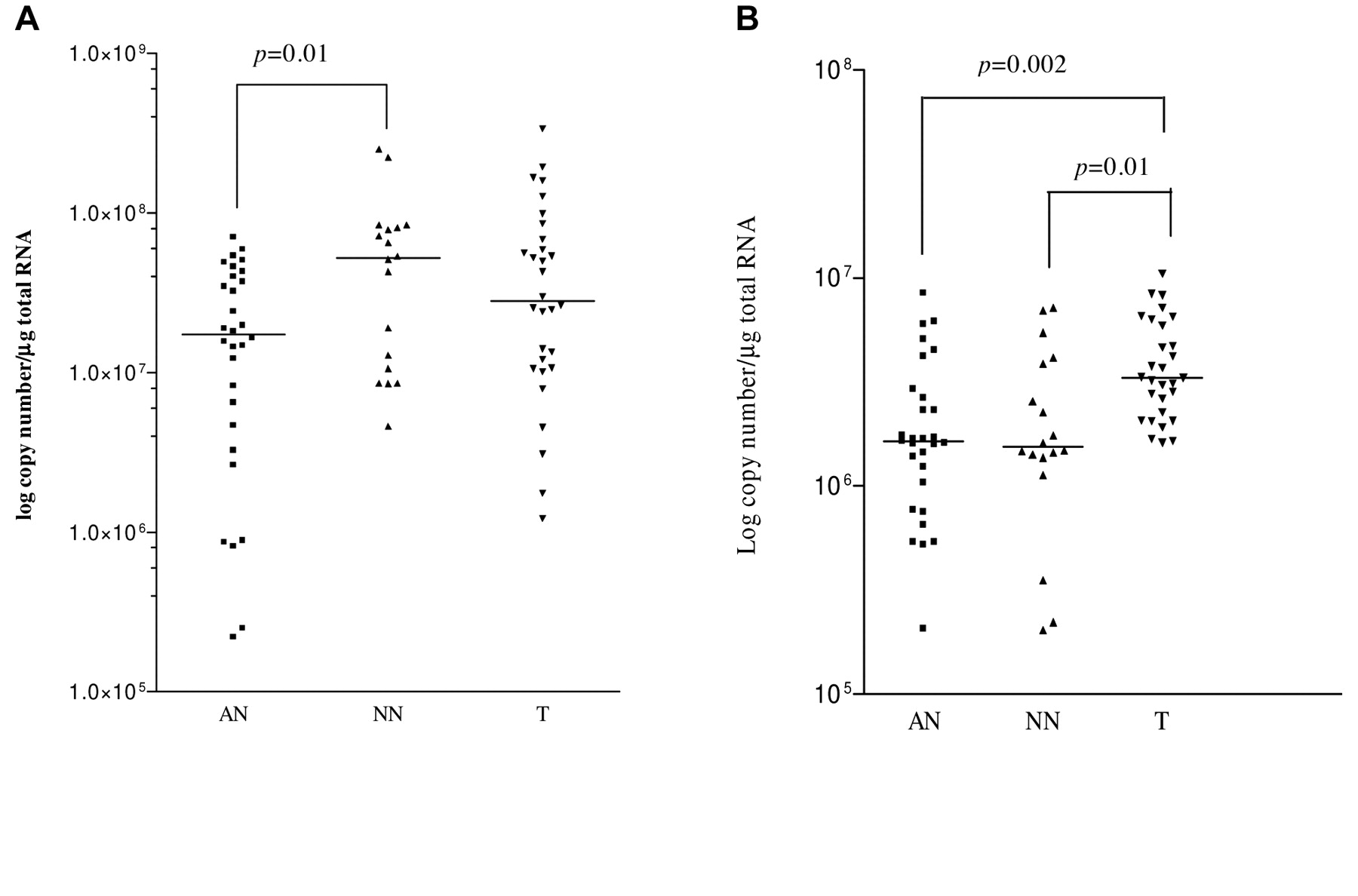
1αOHase and VDR mRNA was detected in all samples. Median 1αOHase mRNA levels were significantly higher (Mann-Whitney U test) in the NN samples (5.2×10⁷; range 4.59×10⁶ - 2.52×10⁸) compared with AN samples (1.7×10⁷; range 1.04×10⁵ - 1.57×10⁸; p=0.01) (Figure 1A). Median 1αOHase mRNA copy number in T samples (2.8×10⁷; range 1.75×10⁶ - 3.38×10⁸) was higher than in paired AN samples (p=0.04) with higher levels (>3 fold) detected in 11/30 tumours and lower levels in 4 tumour samples.

1αOHase (A) and vitamin D receptor (B) mRNA copy numbers in breast tumour (T), adjacent non-cancerous normal tissue (AN) and healthy normal breast tissue (NN). 1αOHase primers and probe were: forward 5′ (GCT ATT GGC GGG AGT GGA C), reverse 5′ (GCC GGG AGA GCT CAT ACA GA) and a hydrolysis probe (CCC AAG AGA GCG TGT TGG ACA CCG). VDR primers and probes (forward 5′-GCTATTGGCGGGAGTGGAC-3′ and reverse 5′-GCCGGGAGAGCTCATACAGA-3′) and a hydrolysis probe (5′ -CCCAAGAGAGCGTGTTGGACACCG-3′).
Median VDR mRNA levels were significantly higher in T samples (3.3×10⁶; range 7.12×10⁴ - 8.7×10⁷) compared to both AN (1.65×10⁶; range 5.21×10⁵ - 8.45×10⁶; p=0.002) and NN samples (median 1.55×10⁶; range 4.83×10⁵ - 7.19×10⁶; p=0.01). There was no difference between AN and NN samples. Immunohistochemistry confirmed VDR protein expression with staining occurring predominantly in the epithelium.
Discussion
The striking observation from these data is the reduced expression of 1αOHase mRNA in healthy tissue from patients with breast cancer compared to that from patients with benign disease. This expression pattern is similar to the one we have previously reported in the colon (2). There are three possible explanations for this finding. First is that it is merely a reflection of age as the samples of normal breast from women without cancer were from younger women than those with cancer. However, there was no correlation between 1αOHase mRNA levels and age in either the normal controls or the adjacent normal breast tissue from cancer cases. Moreover, there was no statistical difference in mRNA levels in the NN samples between younger (<40 years) and older (>40 years) patients. Second, the down-regulation in adjacent healthy tissue may reflect a field effect of the tumour. Our results indicate that this is not the case as 1αOHase mRNA levels were approximately constant between normal breast tissue immediately adjacent and up to 10 cm away from the cancer (Figure 2). A similar result was obtained in normal colonic tissue from patients with colon cancer (unpublished observations). Third, that there is a general mechanism of dysregulation of the vitamin D axis in breast tissue of women who subsequently develop breast cancer. This could result in a predisposition to tumour development given the anti-proliferative and pro-apoptotic effects of active vitamin D (4). Previous work has implicated aberrant promotor methylation as an important mechanism for the development of both breast and colon cancer and hypermethylation of the 1αOHase gene promotor has recently been reported in more than 40% of primary breast cancer (5). Our data imply that such hypermethylation may be present in normal tissue and account for the observed down-regulation. If present, it would suggest that disruption of this hypermethylation might offer a novel chemopreventive approach to breast cancer.
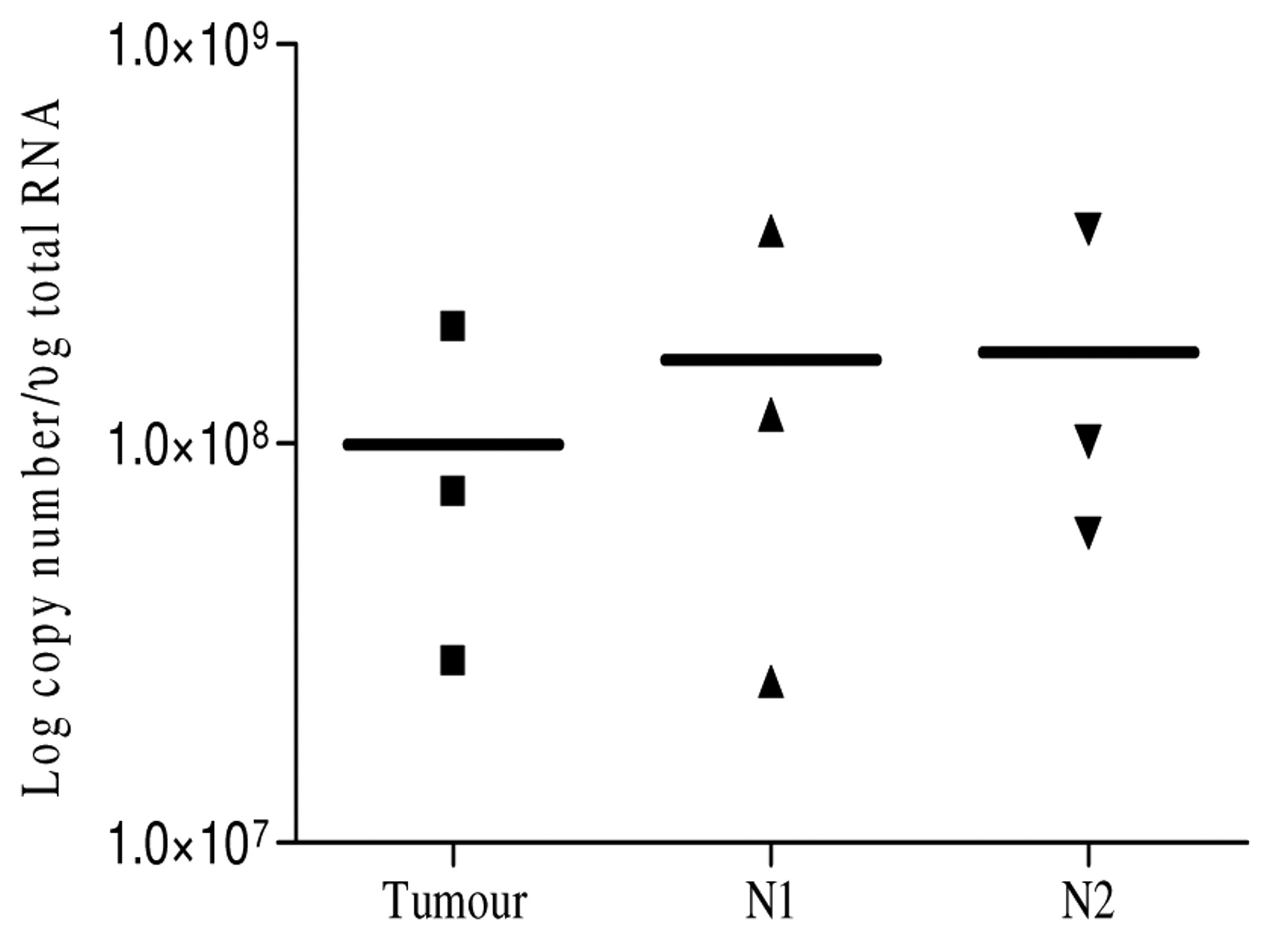
Field effect: 1αOHase mRNA expression in breast tumours (T), normal tissue taken from beside the tumour (N1) and as far away from the tumour as possible (N2).
We did not observe any association between either 1αOHase or VDR mRNA levels and oestrogen receptor status, histological grade of tumour, presence of vascular invasion or lymph node metastases. In contrast, in breast cancer cell lines, expression of 1αOHase has been reported to be higher in well-differentiated, estrogen receptor (ER)-positive cells. In agreement with previous studies, VDR mRNA and protein levels were significantly higher in tumours compared to both adjacent normal and control samples (Figure 1B). The biological significance of this is uncertain.
Conclusion
In conclusion, these early data confirm the importance of the vitamin D axis in breast cancer and suggest that assessment of 1αOHase mRNA levels in women may be useful in assessing risk of future breast cancer development.
Footnotes
- Received August 15, 2008.
- Accepted October 30, 2008.
- Copyright© 2009 International Institute of Anticancer Research (Dr. John G. Delinassios), All rights reserved


