


Abstract
Background/Aim: Fatty liver disease is increasing in the developed and developing world. Liver metastasis from malignant lymphoma in the fatty liver is poorly understood. In a previous report, we developed color-coded imaging of the tumor microenvironment (TME) of the murine EL4-RFP malignant lymphoma during metastasis, including the lung. In the present report, we investigated the potential and microenvironment of the fatty liver induced by a choline-deficient diet as a metastatic site in this mouse lymphoma model. Materials and Methods: C57BL/6-GFP transgenic mice were fed with a choline-deficient diet in order to establish a fatty liver model. EL4-RFP cells were injected in the spleen of normal mice and fatty-liver mice. Metastases in mice with fatty liver or normal liver were imaged with the Olympus SZX7 microscope and the Olympus FV1000 confocal microscope. Results: Metastases of EL4-RFP were observed in the liver, ascites and bone marrow. Primary tumors were imaged in the spleen at the injection site. The fewest metastases were observed in the fatty liver. In addition, the fewest cancer-associated fibroblasts (CAFs) were observed in the fatty liver. Conclusion: The relative metastatic resistance of the fatty liver may be due to the reduced number of CAFs in the fatty livers. The mechanism of the effect of the choline-deficient diet is discussed.
- EL4-RFP
- malignant lymphoma
- liver metastasis
- choline-deficient diet
- fatty liver
- cancer-associated fibroblasts
- methionine-dependence
Non-alcoholic fatty liver disease (NAFLD) has increased in developed countries, with a prevalence of 20-30% in Europe and the United States and 12-30% in Asia (1-8). Obesity is a risk factor for NAFLD and has influenced the increase in NAFLD (9). An important question is whether fatty liver has an influence on cancer metastasis.
We have previously shown that murine EL4-RFP malignant lymphoma cells metastasize to multiple sites including the liver (10, 11). We have previously described color-coded imaging of the tumor microenvironment (TME) of cancer cells (11-20).
A syngeneic color-coded imageable lymphoma model, using the EL4 cell line, was previously developed to visualize recruitment of host stromal cells in the TME during metastasis. Large EL4-RFP liver metastases in C57BL/6-GFP mice contained GFP-expressing stromal cells derived from the host (10). A subsequent study whereby liver metastases were formed after splenic injection of EL4-RFP cells also demonstrated that the liver metastases were rich in stromal cells including macrophages, fibroblasts, dendritic cells and normal lymphocytes (11).
In the present report, the TME of EL4-RFP metastasis in mice with fatty liver and normal liver were compared, with the use of EL4-RFP.
Materials and Methods
GFP transgenic mice. Transgenic C57BL/6-GFP mice were obtained from the Research Institute for Microbial Diseases (Osaka University, Osaka, Japan). The C57BL/6-GFP mice expressed Aequorea victoria GFP under the control of the chicken β-actin promoter and cytomegalovirus enhancer (21). Eight-week-old GFP immunocompetent C57BL/6-GFP transgenic mice were used as the host for RFP-EL4 lymphoma cells. Fatty liver was generated in C57BL/6-GFP mice (n=3) on a choline-deficient diet (CDD) for 4 weeks (22). Another group of mice (n=3) were provided with the control diet (CD) for 4 weeks until the end of the experiment. Diets were purchased from Oriental Yeast Co. Ltd., Tokyo.
Cell line and culture conditions. EL-4, a mouse lymphoma cell line, was previously established from a lymphoma induced in a C57BL/6 mouse by 9, 10-dimethyl-1, 2-benzanthracene (10, 11). The cells were maintained in RPMI 1640 supplemented with 10% heat-inactivated fetal bovine serum (FBS), 1% penicillin and streptomycin (Gibco-BRL, Grand Island, NY). The cells were cultured in a humidified atmosphere containing 5% CO2 at 37°C.
Red fluorescent protein (RFP) transduction of lymphoma cells. EL4 lymphoma cells were labeled with RFP as previously reported using retrovirus-based vector expressing RFP (10, 11, 14, 23-25).
Color-coded lymphoma-host liver model. EL4-RFP mouse malignant lymphoma cancer cells (2.0×10⁶) were injected in the spleen of mice on day 14, after the start of the CDD or CD. All mice were sacrificed on day 28. EL4-RFP lymphoma cells, growing in vitro, were harvested and washed three times with cold serum-free medium and then re-suspended with serum-free RPMI 1640 medium. EL4-RFP lymphoma cells were then injected in the spleen of C57BL/6-GFP transgenic mice. Liver metastases tumors, primary tumors in the spleen, ascites and bone marrow cells were cultured for several weeks from CD and CDD mice (10,11).
Ex vivo imaging. The SZX7 microscope and FV1000 confocal microscope (Olympus Corp. Tokyo, Japan) were used for imaging.
Histology. Tumors were prepared for histological analysis using including hematoxylin and eosin (H&E) staining using previously published protocols (26).
Statistical analysis. All data are expressed as the mean ± SD and analyzed using the two-sided t-test. Differences were considered significant when the two-sided p-value was less than 0.05.
Study approval. All experiments were conducted in accordance with the Institutional Guidelines of Gifu University and were approved by the Animal Research Committee and the Committee on Living Modified Organisms of Gifu University.
Results and Discussion
Establishment of a fatty liver model. To establish a fatty liver mouse model, C57BL/6-GFP mice were fed a choline-deficient diet (CDD) for 4 weeks. Mice were divided into two groups, a CDD group and a control-diet (CD) group. In each group, EL-4-RFP metastases were observed in the liver, ascites and bone marrow, as well as in the spleen at the primary injection site.
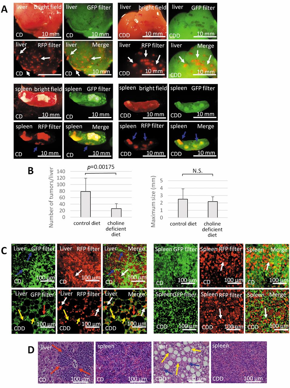
Metastases in normal and fatty liver. Multiple liver metastases and primary tumors in the spleen were observed in each group (Figure 1A). CD mice had more liver metastases than CDD mice (Figure 1B,C). Many lipid droplets were formed in the CDD mice liver (Figure 1C), indicating that liver became fatty on the CDD. H&E staining showed that many lipid droplets occupied the liver tissue (Figure 1D) and fewer cancer cells were observed in the CDD mice, while larger masses of cancer cells were formed in the livers of CD mice (Figure 1A, B). Fewer EL-4 RFP cells and GFP stromal cells, such as fibroblasts were observed in the liver of CDD mice than in CD mice (Figure 1C). On the other hand, in the spleen, there were no significant differences in the tumors size between CD mice and CDD mice (Figure 1C).
Comparison of cells from metastases in fatty and normal liver and other organs cultured in vitro. Liver metastases, primary tumors in the spleen, ascites and bone marrow cells were cultured for several weeks from CD and CDD mice. Phagocytic cells were observed in liver metastases from both CDD and CD mice on day 7 (Figure 2A). Dendritic cells and cancer-associated fibroblasts (CAFs) were also observed in liver metastasis in CD mice, and dendritic cells were observed in CDD mice on day 14 (Figure 2B). CAFs were observed in the spleen of CD and CDD mice (Figure 2C). EL4-RFP lymphoma cells were abundant in ascites (Figure 2D).EL4-RFP cells were observed in the bone-marrow culture (Figure 2E). Bone-marrow cultures contained EL4-RFP cells. Fewer CAFs were observed in the liver metastasis from CDD mice than CD mice (Figure 2F).
It should be noted that CDD-fed rodents have lower methionine levels, as choline is a precursor of methionine (27-38), which may inhibit tumor growth in the liver, due to the enhanced requirement of cancer cells for methionine.
Conclusion
Liver metastasis from colorectal cancer is less frequently observed in patients with hepatic steatosis than in patients with normal liver (21, 30) which may be related to the present study, where an analogous result was obtained with reduction of EL4-RFP metastases in the fatty liver along with fewer CAFs. The fatty liver may not be a suitable environment for CAFs which may be due to re-programmed methionine metabolism which may have occurred during the time the animals were on the CDD (31-46); and as a result, fewer liver metastases were formed.

(A) Bright-field and fluorescence imaging of the liver and spleen. RFP-expressing tumors were observed in the liver and spleen of CD and CDD mice. White arrows indicate liver metastases. Blue arrows indicate primary tumors in the spleen. All images were captured with the Olympus SZX7 microscope. Bar=10 mm. (B) Left bar graphs depict the number of metastases in the liver. Right bar graphs depict the size of the primary tumor. (C) High-magnification images of liver metastasis and primary tumors in the spleen. White arrows indicate EL4-RFP lymphoma cells. Yellow arrows indicate lipid droplets. Red arrows indicate blood cells. All images were captured with the Olympus FV1000 confocal microscope. Bar=100 μm. (D) Histopathological analysis with H&E-stained sections. Red arrows indicate the tumor in the CD mouse liver. Yellow arrows indicate lipid droplets in the CDD mouse liver. Blue arrows indicate lymphoma cells. Bar=100 μm.

High-magnification images of cells cultured from tumors and metastases, which were harvested from the CD and CDD mice. Images were obtained with the Olympus FV1000 confocal microscope. CD=control diet CDD=choline deficient diet. Bar=100 μm. (A) Cultures of liver metastases in CD and CDD mice on day 7. Yellow arrows indicate phagocytosis. (B) Cultures of liver metastases from CD and CDD mice on day 14. White arrows indicate EL4-RFP cells. Blue arrows indicate cancer-associated fibroblasts (CAFs). Yellow arrows indicate dendritic cells. (C) Cultures of EL4-RFP lymphoma from the spleen of CD and CDD mice on day 7. Yellow arrows indicate CAFs. (D) Cultures of EL-4-RFP lymphoma ascites from CD and CDD mice on day 7. Red arrows indicate blood cells. Yellow arrows indicate phagocytosis. (E) Cultures of bone marrow from CD and CDD mice on day 7 after EL-4-RFP implantation. Abundant EL4-RFP lymphoma cells and stromal cells expressing GFP were observed. (F) The number of CAFs in the liver was counted. Values are expressed as mean±SD.
Footnotes
This article is freely accessible online.
Conflicts of Interest
None of the authors have any conflict of interest in regard to this study.
- Received April 21, 2017.
- Revision received May 18, 2017.
- Accepted May 24, 2017.
- Copyright© 2017, International Institute of Anticancer Research (Dr. George J. Delinasios), All rights reserved


