


Abstract
Background/Aim: Multiple myeloma is still an incurable hematological malignancy of monoclonal B-lymphocytes. While standard chemotherapy regimens have been used for years, novel agents like lenalidomide and bortezomib have become an essential part of today's therapies and significantly improve therapeutic efficacy. Nevertheless, new therapeutic strategies are still indispensable. Aberrant activation of Wnt/β-catenin signaling promotes development of several cancers. Recently, it has been demonstrated that the Wnt pathway is activated in both lymphoma and myeloma. Thus, Wnt signaling molecules are attractive candidates for the development of new targeted-therapies. Naftifine was used in the present study since it has chemical features similar to those of other known WNT inhibitors. Materials and Methods: The anti-tumor apoptotic effect of naftifine at doses ranging from 0.1-200 μM was investigated on two human and one murine lymphoma, as well as in one murine and three human myeloma cell lines, and determinded by the 3-(4,5-dimethylthiazol-2-yl)-2,5-diphenyltetrazolium-bromide (MTT) assay. Results: Naftifine significantly reduced cell viability in all tested myeloma and lymphoma cell lines in a dose-dependent manner, while healthy cells were only slightly affected. Conclusion: Naftifine exhibits toxicity to hematological neoplasms in vitro.
Multiple myeloma (MM) represents a hematological neoplasm of post-germinal center B-lymphocytes, that is characterized by the accumulation of malignant secretory plasma cells in the bone marrow and mostly occurs with monoclonal protein in either peripheral blood and/or urine. Heterogeneous and unspecific clinical symptoms often delay early diagnosis particularly during the onset of the disease (1). MM is primarily diagnosed in elderly patients with a median age at diagnosis of 69 years (2). Over the last decade, newly-introduced therapeutic regimens including Immunomodulatory Drugs (IMiDs) like bortezomib, lenalidomide and thalidomide significantly improved treatment outcome and patient survival. Despite this major progress in the treatment of MM most patients might eventually relapse, underlining the need for new therapeutic approaches.
Various signaling pathways involved in cancerogenesis represent promising targets in cancer therapy since they have been shown to be involved in apoptosis induction, differentiation and regulation of cell proliferation. Of particular interest is aberrant signaling of the Wnt pathway as it has been shown to induce and maintain major oncogenic effects (3-7). In this context, exaggerated Wnt signaling is well-documented for oncogenes as well as the tumoural course of lymphoma and MM (8-12). Specific inhibition of Wnt signaling results in suppressed progression of lymphoma and MM, thus influencing the Wnt signaling cascade might represent a valuable therapeutic approach (12, 13).
In our previous studies we confirmed that ethacrynic acid (EA), ciclopiroxolamine (CIC), piceatannol (PIC) and piroctoneolamine (PO) target Wnt/beta-catenin signaling molecules and might be effective in the therapy of various, especially hematopoietic types of cancer (14-24). Additionally, subsequent in-vitro studies showed that cinnarizine and flunarizine, chemically related agents to other known Wnt inhibitors, were also selective inducers of apoptosis in hematologic malignancies (25, 26).
Concerning its chemical features, naftifine is closely related to terbinafine, which has demonstrated anti-carcinogenic properties, and is distantly related to other known Wnt inhibitors. For this reason, we investigated whether naftifine could also exhibit any cytotoxic effects on both myeloma and lymphoma cells and demonstrated for the first time that naftifine is effective in the treatment of myeloma and lymphoma cells in vitro.
Materials and Methods
Cell lines and culture conditions. Cell lines were obtained from DSMZ (Braunschweig, Germany) or ATCC (LGC Standards, Wesel, Germany) and incubated at 37°C with 5% CO2 and at 90% humidity.
The human myeloma cell lines KMS 18, RPMI-8226 and U-266 (all obtained from DMSZ, Braunschweig, Germany) were cultured in RPMI-1640 medium (PAA, Pasching, Austria), supplemented with 5% of heat-inactivated fetal calf serum FCS (Invitrogen, Darmstadt, Germany) and 1% penicillin/ streptomycin (Seromed, Jülich, Germany). The human lymphoma cell lines Raji and SU DHL 4 were cultured under identical conditions as human myeloma cell lines. MPC-11 and RAW 264,7 (ATCC, LGC Standards GmbH, Wesel, Germany) are both murine cell lines. MPC-11 is a murine plasmocytoma cell line and RAW 264,7 is a leukaemic monocyte macrophage cell line. Cells were cultured in RPMI-1640 medium supplemented with 5% of heat-inactivated FCS and 1% penicillin/ streptomycin. RAW 264,7 cells were harvested by using 0.05% trypsin-EDTA solution (Invitrogen, Darmstadt, Germany).
The human colon fibroblast cell line CCD-18Co was obtained from ATCC (LGC Standards, Wesel, Germany) and cultured in ATCC-formulated Eagle's Minimum Essential Medium (LGC Standards, Wesel, Germany) supplemented with 15% of heat-inactivated FCS and 1% penicillin/ streptomycin. Cells were harvested by 0.05% trypsin-EDTA solution (Invitrogen, Darmstadt, Germany), centrifuged at 1,200 xg for 7 min and re-suspended in 1mL media to define the cell count. The medium was renewed every 3 days.
Drugs and chemical reagents. Naftifine was the drug under investigation in this study. Naftifine was purchased from Sigma-Aldrich (Steinheim, Germany). Naftifine was tested at concentrations ranging from 0.1 to 200 μM for 72 h.
Cell viability assay with 3-(4,5-dimethylthiazol-2-yl)-2,5-diphenyltetrazolium-bromide (MTT). The effectiveness of naftifine was determined by cell viability in MTT assay. Viable cells convert the yellow MTT (Sigma Aldrich, Steinheim, Germany) into purple formazan when taken-up into the mitochondria. Cells were plated at 1×10⁴ well/ 100μL in 96-well plates, left to adhere overnight in the incubator. The next day media was removed and renewed containing various concentrations of Naftifine. After 21 h 1 μL MTT (5mg/ml) was added to each well and incubated for another 3 h. Then 80 μL of the media were removed and 50 μL of acidified isopropanol was added for cell lysis. After shaking for 10 min the amount of formazan was measured at 565nM. The measured amount of formazan in treated cells was compared to untreated cells.
Statistical analysis. Values are given as mean±standard deviation (SD). At least three separate and independent experiments were performed with each cell line. Two-tailed Student's t-test was used for statistical analysis. A p-value less than 0.05 was considered significant.
Inhibitory concentration (IC50) of naftifine for human and murine lymphoma, multiple myeloma and control cell lines.
Results
Titration of naftifine. As a first step, we determined optimal concentrations of naftifine leading to significant decrease in viability of all tested myeloma and lymphoma cells. As healthy controls CCD-18Co colonic fibroblasts were investigated by MTT assays. The mean 50% inhibitory concentration (IC50) after 72 h was calculated following titration. IC50 values of naftifine employed after 72 h of incubation are shown in Table I.
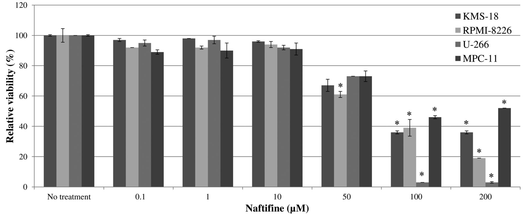
Effect of naftifine on viability of human myeloma cells. The viability of all tested human myeloma cells was affected by naftifine. Administered concentrations of naftifine starting from 50 μM significantly decreased the viability of myeloma cells in a concentration-dependent manner. Maximum efficiency was observed with concentrations higher than 50 μM. Results were as shown in Figure 1.
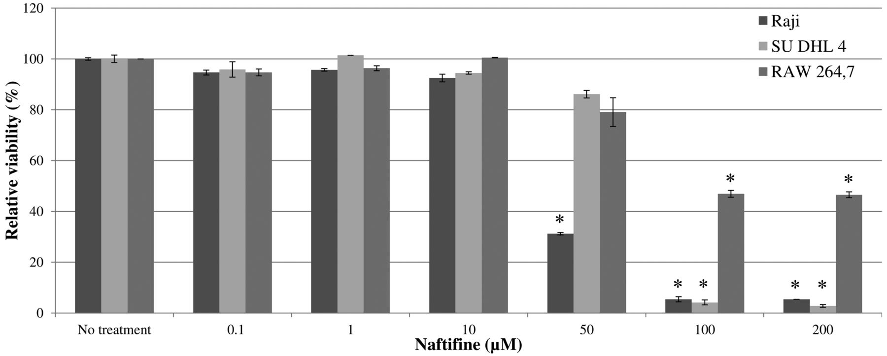
Effect of naftifine on viability of human lymphoma cells. Exposure to naftifine concentrations starting from 50 μM also significantly decreased lymphoma cell viability in all tested cell lines. The IC50 of Raji cells was attained after treatment with 45 μM. SU DHL 4 lymphoma cells were less susceptible to the toxicity of naftifine. At least 72 μM naftifine were required to decrease their viability to a level of 50%. Figure 2 presents the respective results.
Effect of naftifine on viability of murine myeloma cells and murine macrophages. The effects of naftifine treatment in human myeloma and lymphoma cells were reproducible in murine myeloma cells as well as in leukaemic monocyte macrophages and are presented in Figures 1 and 2. The concentrations required to significantly decrease viability in MPC-11 myeloma cells were slightly higher compared to those for human myeloma cells. Naftifine at 90 μM sufficiently reduced the vitality to approximately 50%. Additionally, RAW 264,7 monocytes were investigated to determine if naftifine exhibits any toxicity towards myeloid cells. A significant influence on the viability of RAW 264,7 cells occurred at comparable concentrations as for MPC-11 cells.

Effect of naftifine on viability of KMS-18, RPMI-8226, U-266 and MPC-11 human and murine myeloma cells. Cells were cultured with naftifine for 72 h. Viability was measured by the 3-(4,5-dimethylthiazol-2-yl)-2,5-diphenyltetrazolium-bromide (MTT) assay. Results represent data from three independent experiments. Data are the mean±SD. *p<0.05 compared to untreated cells.

Effect of naftfine on viability of Raji, SU DHL 4 and RAW 264,7 human lymphoma and murine leukaemic monocyte macrophage cells. Cells were cultured with naftfine for 72 h. Viability was measured by the 3-(4,5-dimethylthiazol-2-yl)-2,5-diphenyltetrazolium-bromide (MTT) assay. Results represent data from three separate experiments each. Data are shown as mean±SD.*p<0.05 compared to untreated cells.
Effect of naftifine on viability of healthy controls. We chose CCD-18Co colon fibroblasts in order to analyze the toxicity of naftifine towards healthy cells. CCD18-Co cells tolerated high doses of naftifine as concentrations of more than 100 μM were needed for a significant viability reduction. Results are shown in Figure 3.

Effect of naftifine on viability of CCD18-Co cells which served as controls. Cells were cultured with naftifine for 72 hours. Viability was measured by the 3-(4,5-dimethylthiazol-2-yl)-2,5-diphenyltetrazolium-bromide (MTT) assay. Results represent data from three separate experiments each. Data are shown as mean±SD. *p<0.05 compared to untreated cells.
Discussion
MM represents a malignant neoplasm of plasma cells caused by frequent gene mutations with or without chromosomal translocations (27). Currently, treatment is characterized by a primarily initiated high-dose therapy with chemotherapeutics, optionally followed by hematopoietic stem cell transplantation (27-30). Over the last decades, numerous innovations have been achieved in the development and clinical use of innovative therapeutic agents like IMiDs and their transition into clinical practice (31, 32). Despite these treatment innovations MM currently remains incurable in patients solely treated with chemotherapy(3).
In this context, targeting the canonical Wnt pathway might be an interesting approach as Wnt signaling represents an excellent example of abrogated signaling pathways in MM (8-12). Development and proliferation of MM cells is, among others, dependent on the bone marrow microenvironment, wherein bone marrow stromal cells enhance Wnt signaling by the release of Wnt ligands, consequently leading to an enhanced proliferation activity of MM cells (32-34). As a consequence, the inhibition of Wnt/β-catenin signaling suppresses MM growth as shown in recent in-vitro and in-vivo studies (35). Thus, the Wnt signaling pathway emerged as an attractive therapeutic target for MM.
Recently, our workgroup revealed four drugs, PIC, EA, CIC and PO to be efficient inducers of apoptosis in lymphoma and myeloma cells in vitro. The latter three having already proved their efficacy in subsequent in vivo studies. They significantly reduced the tumor growth and prolonged the overall survival in myeloma-bearing mice. All four drugs rendered the tested cell lines more sensitive to other agents and influenced the Wnt pathway through targeting either β-catenin itself or its downstream factors (14-24, 35).
These promising effects on both cancer cell survival and Wnt signaling encouraged us to determine whether naftifine, which is distantly related to other known Wnt inhibitors, displays any cytotoxicity towards MM and lymphoma cells. Recent in vitro and in vivo studies have shown terbinafine, which is a chemical analogue of naftifine, to possess various anti-carcinogenic properties. Terbinafine dose-dependently decreased cell viability of different human malignant cells by interrupting the cell cycle at the G0/G1 transition and inhibiting the cyclin-dependent kinase system as the levels of p53, p21/Cip1 and p27/Kip1 proteins were augmented. Additionally, the binding between p53 protein and p53 consensus binding site in p21/Cip1 promoter DNA probe was increased, suggesting an involvement of the p53-associated signaling pathway. In a subsequent in vivo setting, terbinafine injections led to obvious decline in tumor size (36). In another recent in vitro study of human oral squamous cell carcinoma cells, terbinafine reduced proliferation and induced apoptosis by suppression of the Raf-MEK-ERK pathway (37).
Naftifine is a synthetic, broad-spectrum anti-fungal agent, used topically to treat superficial dermatomycoses such as tinea pedis, tinea cruris, and tinea corporis. It is an allylamine compound with antifungal and antimycotic activities by inhibiting fungal squalene epoxidase and preventing ergosterol biosynthesis. Naftifine also showed antibiotic activity against Gram-positive as well as Gram-negative bacteria and also demonstrated anti-inflammatory properties, such as a reduction in superoxide production and a reduction in polymorphonuclear leukocyte chemotaxis/ endothelial adhesion (38-40).
We found only few studies investigating other possible qualities of naftifine beyond the ones described above. Its utilization in oncology and its potential influence on neoplasia was not so far addressed, particularly in the light of the promising in vitro and in vivo results with its chemical analogue terbinafine. However, our data also indicate that naftifine might have an impact on the proliferation capacity of hematological malignancies. We showed that naftifine significantly reduced the viability of all tested myeloma and lymphoma cell lines. Both human and murine cells were equally affected by naftifine in a dose-dependent manner. However, slightly higher concentrations were needed in murine myeloma cells. Doses of approximately 75 μM decreased cell viability by 50% in most myeloma and lymphoma cell lines, but Raji lymphoma cells were more sensitive to naftifine. Most interestingly, CCD-18Co colonic fibroblasts, that served as healthy controls, tolerated higher doses of naftifine as doses lower than 200 μM did not significantly influence viability, thus suggesting a favorable tolerability of naftifine concerning healthy tissues.
Owing to its chemical relationship to other known Wnt inhibitors, naftifine might also interfere with signaling molecules embedded in the Wnt and/or associated signaling pathways. However, further studies are required in this respect to determine where an intereference in the Wnt signaling pathway exactly takes place.
Overall, naftifine revealed cytotoxic potential towards MM and lymphoma cells and hardly altered the viability of healthy controls. It might, therefore, have the potential to serve as a future therapeutic tool in the treatment of hematological malignancies. Further in vitro experiments are warranted to provide deeper insight over the molecular mechanisms and to pave the way for future in vivo studies.
Acknowledgements
L.C. Schmeel and F.C. Schmeel contributed equally to this study and are both first authors.
Footnotes
↵* These Authors contributed equally to this study.
- Received June 16, 2015.
- Revision received July 22, 2015.
- Accepted August 7, 2015.
- Copyright© 2015 International Institute of Anticancer Research (Dr. John G. Delinassios), All rights reserved


