


Abstract
Background: In tumors, monocytes differentiate into tumor-associated macrophages following interaction with cancer cells. We have previously reported that angiogenesis- and chemotaxis-related factors are associated with human monocyte differentiation following interaction with colon cancer cells. However, the exact nature of factors remains unknown. We investigated factors associated with differentiation of human colon cancer cells following interaction with monocytes. Materials and Methods: The human colon cancer cell line DLD-1 was co-cultured with the human monocyte cell line THP-1. mRNA expression was analyzed by quantitative real-time PCR. Results: Expression of interleukin-1β, matrix metalloproteinase (MMP)-1, MMP-2, and MMP-3 increased in human colon cancer cells after co-culture with monocytes. Conversely, the expression of monocyte chemotactic protein-1, tumor necrosis factor-α, and signal transducer and activator of transcription-3 did not increase. Conclusion: Differentiation of human colon cancer cells following interaction with monocytes may be associated with angiogenesis and metastasis but not chemotaxis and signaling pathways. Thus, angiogenesis- and metastasis-related factors associated with differentiation of human colon cancer cells may constitute important targets for colon cancer therapy.
Monocytes originate from the bone marrow. They develop and are released into the peripheral circulation. They then enter and populate tissues as macrophages. Subpopulations of macrophages exist, and are defined by anatomical location and functional phenotype (1, 2).
Recently, it was established that most solid tumors were abundantly populated with macrophages, termed tumor-associated macrophages. Tumor-associated macrophages are considered to exert antitumor effects and promote malignant progression (3-11). Several reports describe an association between macrophage number and prognosis in various types of human cancers. Extensive tumor-associated macrophage infiltration is associated with poor prognosis in breast, cervical, and bladder cancer; conversely, their presence is associated with a good prognosis in stomach, lung, and colorectal cancer (4, 6, 7, 12). These discrepancies suggest that monocytes differentiate into tumor-associated macrophages following interaction with cancer cells and that their character differs depending on the cancer type. We previously reported that angiogenesis- and chemotaxis-related factors were associated with differentiation of human monocytes into tumor-associated macrophages following interaction with colon cancer cells (13). However, the nature of these factors remains unknown. We examined changes in mRNA expression of human colon cancer cells following interaction with monocytes to identify factors associated with their differentiation.
Materials and Methods
Cells. DLD-1 cells obtained from the Japan Health Sciences Foundation (Tokyo, Japan), and THP-1 cells obtained from DS Pharma Biomedical (Osaka, Japan) were cultured in a 5% CO2 atmosphere at 37°C in RPMI-1640 medium (Wako Pure Chemical Industries, Ltd., Osaka, Japan) containing 10% fetal calf serum supplemented with 100 units/ml each of penicillin and streptomycin (Wako Pure Chemical Industries, Ltd.).
Cell co-culture. DLD-1 and THP-1 cells were co-cultured using a cell culture insert (Becton, Dickinson and Co., Franklin Lakes, NJ, USA) with a 0.4-μm porous membrane to separate the upper and lower chambers. DLD-1 cells were cultured in the upper chamber at 2×10⁵ cells/ml, and THP-1 cells were cultured in the lower chamber at 2×10⁵ cells/ml. DLD-1 and THP-1 cells were collected 0, 1, 3, and 5 days after co-culture.
Primer sequences for real-time PCR.
RNA extraction. Total RNA extractions from DLD-1 and THP-1 cells were performed using TRIzol Reagent (Invitrogen Corporation, Carlsbad, CA, USA) according to the manufacturer's protocol. RNA was quantified by absorbance at 260 nm. cDNA was synthesized using reverse transcriptase with Oligo(dT)20 (Toyobo Co., Ltd., Osaka, Japan).
Quantitative real-time PCR. mRNA expression was analyzed by quantitative real-time PCR (Model MiniOpticon; Bio-Rad Laboratories, Inc., Hercules, CA, USA) using SsoFast EvaGreen Supermix (Bio-Rad Laboratories, Inc.). After initial heat denaturation at 95°C for 3 min, PCR conditions were set at 95°C for 10 s and 60°C for 30 s for 40 cycles. The primers used are shown in Table I. Relative quantifications were achieved by normalization to the value of the housekeeping gene β-actin. Data are expressed as change (n-fold) in mRNA expression compared with co-culture at day 0.
Results
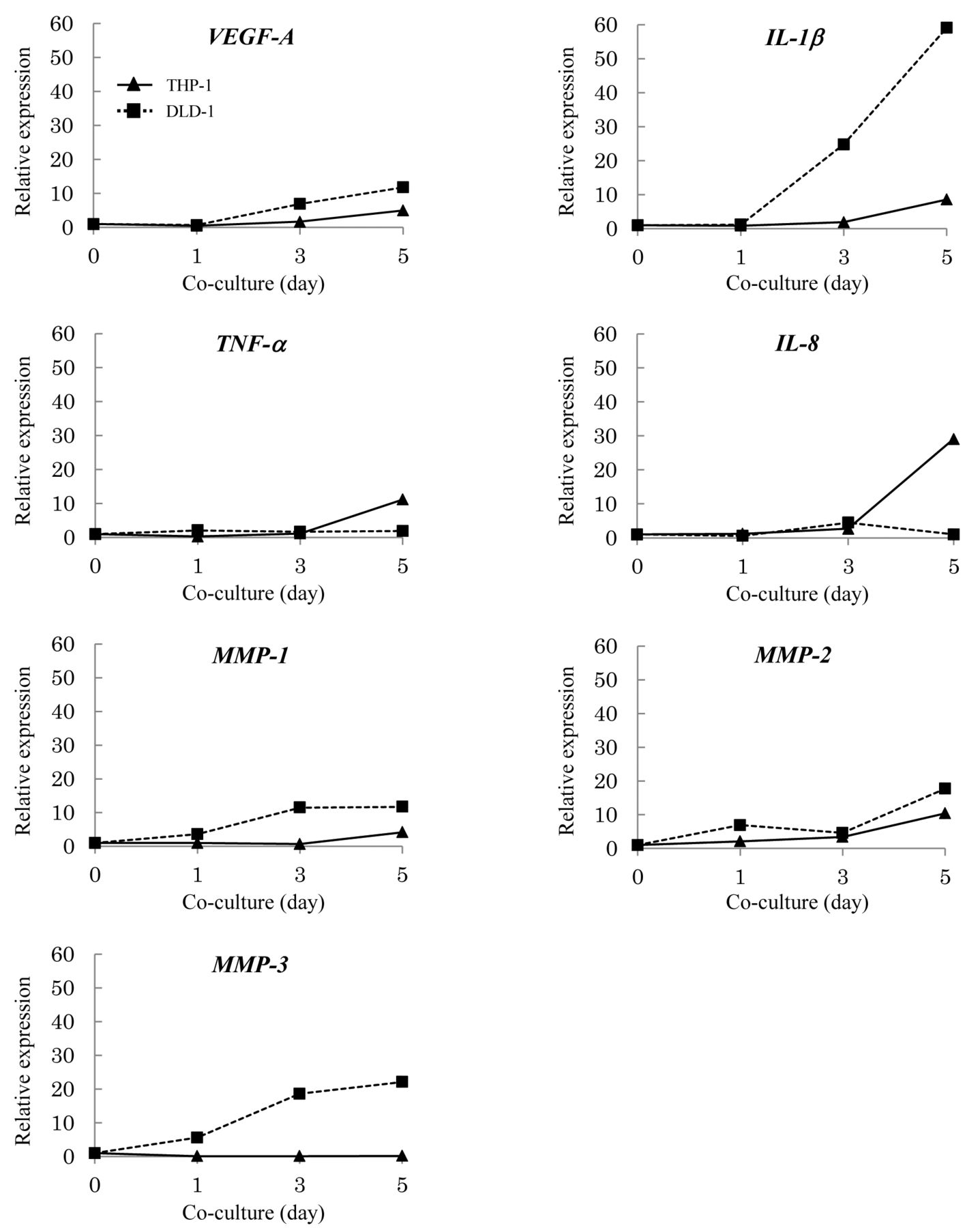
mRNA expression of angiogenesis- and metastasis-related factors in human colon cancer cells is increased following co-culture with monocytes. mRNA expression in DLD-1 cells after co-culture with THP-1 cells was analyzed by quantitative real-time PCR to identify factors associated with human colon cancer cell differentiation. Factors associated with human monocyte differentiation are known to function in tumor progression (14-19). We previously reported that mRNA expression of angiogenesis-related factors such as vascular endothelial growth factor (VEGF)-A and interleukin (IL)-8 increased and that of signaling pathway-related factors such as nuclear factor (NF)-κB did not increase in DLD-1 cells after co-culture with THP-1 cells (Table II) (13). In addition to identifying increased expression of chemotaxis-, angiogenesis-, metastasis-, and signaling pathway-related factors, we quantitatively evaluated mRNA expression in DLD-1 cells co-cultured with THP-1 cells. IL-1β mRNA expression increased 24.8-fold by day 3 and 59.1-fold by day 5; matrix metalloproteinase (MMP)-1 mRNA expression increased 11.5-fold by day 3 and 11.7-fold by day 5; MMP-2 mRNA expression increased 17.7-fold by day 5; and MMP-3 mRNA expression increased 18.6-fold by day 3 and 22.1-fold by day 5. This indicates that mRNA expression of IL-1β, MMP-1, MMP-2, and MMP-3 in DLD-1 cells increased after co-culture. Furthermore, the mRNA expression of IL-1β, MMP-1, and MMP-3 in DLD-1 cells increased in a time-dependent manner after co-culture. mRNA expression of MMP-7 and membrane type 1 (MT1)-MMP also demonstrated an increasing trend in DLD-1 cells co-cultured with THP-1 cells (Table II). Conversely, mRNA expression of monocyte chemotactic protein (MCP)-1, tumor necrosis factor (TNF)-α, and signal transducer and activator of transcription-3 (STAT3) did not increase until day 5 in DLD-1 cells co-cultured with THP-1 cells (Table II). Therefore, it is clear that mRNA expression in human colon cancer cells changed following interaction with monocytes. Our results suggest that human colon cancer cell differentiation following interaction with monocytes is associated with angiogenesis and metastasis but not chemotaxis and signaling pathways.
mRNA expression in DLD-1 cells following co-culture with THP-1 cells.

Differential expression of angiogenesis- and metastasis-related factors in DLD-1 and THP-1 cells following co-culture. mRNA expression after co-culture was analyzed by quantitative real-time PCR. Relative quantifications were achieved by normalization to the value of the housekeeping gene β-actin. Data are expressed as change (n-fold) in mRNA expression compared with co-culture at day 0.

mRNA expression of nuclear factor (NF)-κB and signal transducer and activator of transcription-3 (STAT3) in DLD-1 and THP-1 cells, following co-culture. mRNA expression after co-culture was analyzed by quantitative real-time PCR. Relative quantifications were achieved by normalization to the value of the housekeeping gene β-actin. Data are expressed as change (n-fold) in mRNA expression compared with co-culture at day 0.
mRNA expression pattern of chemotaxis-, angiogenesis-, and metastasis-related factors in human colon cancer cells is different from that in monocytes following co-culture. We examined the mRNA expression patterns of factors associated with cell differentiation in DLD-1 and THP-1 cells after co-culture. In DLD-1 cells, mRNA expression of VEGF-A, IL-1β, MMP-1, MMP-2, and MMP-3 increased by day 5 after co-culture, whereas that of TNF-α and IL-8 did not increase until day 5 after co-culture. Conversely, in THP-1 cells, mRNA expression of VEGF-A, IL-1β, TNF-α, IL-8, and MMP-2 increased by day 5 after co-culture, whereas that of MMP-3 did not increase until day 5 after co-culture. mRNA expression of VEGF-A, IL-1β, MMP-1, MMP-2, and MMP-3 increased more in DLD-1 cells after co-culture than in THP-1 cells (Figure 1). It is established that VEGF-A, IL-1β, TNF-α, and IL-8 are angiogenesis-related factors and that MMPs are metastasis-related factors. Our results indicate that the alterations in angiogenesis- and metastasis-related factors in human colon cancer cells after co-culture differ from those altered in monocytes. Furthermore, the mRNA expression patterns of angiogenesis- and metastasis-related factors in human colon cancer cells after co-culture differ from those in monocytes. Therefore, our results suggest that the mechanism of human colon cancer cell differentiation following interaction differs from that of monocyte differentiation.
mRNA expression of signaling pathway-related factors is not increased in human colon cancer cells and monocytes following co-culture. We examined the mRNA expression of signaling pathway-related factors in DLD-1 and THP-1 cells to understand their mechanisms of differentiation following interaction. NF-κB and STAT3 mRNA expressions in DLD-1 and THP-1 cells did not increase up to day 5 after co-culture (Figure 2). This suggests that the mRNA expression of signaling pathway-related factors does not increase in human colon cancer cells and monocytes following interaction.
Reportedly, NF-κB and STAT3 are expressed in many solid tumors and are activated by inflammatory cytokines, such as TNF-α. Therefore, we examined TNF-α mRNA expression in DLD-1 cells after co-culture. TNF-α mRNA expression in DLD-1 cells did not increase until day 5 after co-culture (Figure 1). This suggests that the mechanism of cancer cell differentiation following interaction with monocytes differs depending on cancer type.
Discussion
We showed that mRNA expression of VEGF-A, IL-1β, MMP-1, MMP-2, and MMP-3 increased in human colon cancer cells following co-culture with monocytes. Moreover, we demonstrated that mRNA expression of VEGF-A, IL-1β, TNF-α, IL-8, and MMP-2 increased in human monocytes following co-culture with colon cancer cells. These results indicate that mRNA expression changes following co-culture in both human monocytes and colon cancer cells. Furthermore, our results imply that human colon cancer cell differentiation following interaction is associated with angiogenesis and metastasis but not chemotaxis and signaling pathways. Therefore, human colon cancer cells may promote angiogenesis and metastasis in tumor tissues following interaction with monocytes. Thus, factors associated with human colon cancer cell differentiation may constitute important targets for colon cancer therapy.
Our results also indicate that the alterations in angiogenesis- and metastasis-related factors in human colon cancer cells following interaction differ from those altered in monocytes. In addition, we showed that the mRNA expression patterns of angiogenesis- and metastasis-related factors in human colon cancer cells following interaction differ from those in monocytes. Therefore, the mechanism of differentiation of human colon cancer cells following interaction may differ from that of monocytes. Thus, our results suggest that a synergistic effect, resulting from the interaction between human colon cancer cells and monocytes, promotes the progression of tumors to malignancy. mRNA expression of MMP-1, MMP-2, and MMP-3 in human colon cancer cells and that of MMP-2 in monocytes increases following interaction. It is established that MMPs are associated with the invasive properties of cancer cells (19). Therefore, it is possible that inhibition of MMP expression in human colon cancer cells and monocytes correlates with suppression of tumor infiltration and metastasis. Thus, inhibitors of MMP expression may become targets for colon cancer therapy.
It is known that activated NF-κB and STAT3 are required for the development of colitis-associated cancer. Indeed, NF-κB and STAT3 overexpression at tumor sites may be involved in the differentiation of monocytes into tumor-associated macrophages (20-26). NF-κB and STAT3 are known to be activated by inflammatory cytokines, such as TNF-α. However, this study indicated that NF-κB and STAT3 mRNA expression in DLD-1 and THP-1 cells did not increase until day 5 after co-culture. We also showed that TNF-α mRNA expression did not increase in DLD-1 cells until day 5 after co-culture. Extensive tumor-associated macrophage infiltration reportedly correlates with poor prognosis in breast, cervical, and bladder cancer, whereas the presence of tumor-associated macrophages reportedly correlates with a good prognosis in stomach, lung, and colorectal cancer (4, 6, 7, 12). Thus, our results suggest that the mechanism of cancer cell differentiation following interaction with monocytes differs depending on cancer type.
- Received April 4, 2013.
- Revision received June 3, 2013.
- Accepted June 4, 2013.
- Copyright© 2013 International Institute of Anticancer Research (Dr. John G. Delinassios), All rights reserved
References
In this issue


Jump to section
Related Articles
Cited By...
- Gene Expression in Lipopolysaccharide-treated Human Monocytes Following Interaction with Hepatic Cancer Cells
- Molecular Response of Human Monocytes Following Interaction with Colon Cancer Cells by Pre-treatment with Low-dose Lipopolysaccharide
- Expression of Chemotaxis- and Angiogenesis-related Factors in Human Monocytes Following Interaction with Colon Cancer Cells Is Suppressed by Low-dose Lipopolysaccharide