


Abstract
Elevated serum interleukin-6 (IL-6) levels have been associated with tumor progression and poor prognosis in patients with esophageal carcinoma. The purpose of the present study was to clarify such a relationship in patients with esophageal squamous cell carcinoma with a focus on the possible influence of chemoradiotherapy (CRT) on tumor IL-6 expression. Data regarding 41 patients with clinical T3-T4 tumors who underwent induction chemoradiotherapy followed by surgery (CRT group) and 60 patients with clinical T1-T4 tumors who underwent surgery alone (Surgery group) between 2001 and 2010, were retrospectively analyzed. Tumor IL-6 expression in resected specimens was evaluated by immunohistochemistry. Tumor IL-6 expression was detected in patients with advanced tumors in the Surgery group (21.1% in pstage III-IV vs. 0.2% in pstage I-II; 27.8% in pT3-4 vs. 0% in pT1-2), and also correlated with primary tumor progression and surgical curability in the Surgery group. In addition, patients with IL-6-positive tumors had significantly shorter overall survival than those with IL-6-negative tumors in the CRT group, and tumor IL-6 expression had an independent prognostic value in multivariate analysis, whereas no significant difference in overall survival was observed between patients with IL-6-positive and those with IL-6-negative tumors in the Surgery group. These results indicate that pre-treatment tumor IL-6 expression correlates with primary tumor progression, and CRT-induced tumor IL-6 expression predicts poor prognosis.
Esophageal carcinoma is one of the most malignant tumor types due to its aggressive behavior. Patients with locally advanced tumors have a poor prognosis when treated exclusively by surgical resection. Currently, neoadjuvant chemoradiotherapy (CRT) plays an important role in the multimodal treatment of esophageal carcinoma. Because of the variance in response to CRT, only patients with major histopathological responses will ultimately benefit from this treatment (1). Accordingly, it is important to search for biomarkers that identify sub-groups of patients who may be sensitive to CRT.
Interleukin 6 (IL-6), a potent pro-inflammatory cytokine, has been associated with disease progression in various malignancies (2). Serum IL-6 levels have been shown to be higher in patients with esophageal carcinoma than in healthy controls, and have been correlated with disease progression and poor prognosis (3, 4). IL-6 levels have also been shown to be higher in tumor tissues than in adjacent normal esophageal tissues (4-6). This indicates that IL-6 in tumor tissues plays a pivotal role in the pathological behavior of esophageal carcinoma. In this regard, our previous study demonstrated that elevated serum C-reactive protein and IL-6 levels after CRT were associated with resistance to CRT and poor prognosis in patients with advanced esophageal squamous cell carcinoma (ESCC) who underwent induction CRT, followed by surgery (7). In that study, IL-6 expression was detected within tumor tissues after CRT; however, the relationship between tumor IL-6 expression and patient outcomes remain unclear.
The aim of the present study was to assess tumor IL-6 expression by immunohistochemistry in resected specimens from patients with ESCC who underwent surgery with or without preoperative CRT. The correlation of tumor IL-6 expression was analyzed with clinicopathological parameters and overall survival and the results were compared between patients who underwent surgery with and without pre-operative CRT.
Patients and Methods
Patients. Data regarding 41 patients with clinical T3-T4 esophageal squamous cell carcinoma, who underwent induction CRT followed by esophagectomy (CRT group) and 60 patients with clinical T1-T4 esophageal squamous cell carcinoma, who underwent esophagectomy without any preoperative therapy (Surgery group) at Kyoto Prefectural University of Medicine Hospital between 2001 and 2010 were analyzed in this retrospective study. An immunohistochemical study for tumor IL-6 expression was performed using resected specimens from consecutive patients between 2001 and 2009 in the CRT group, and those from consecutive patients between 2006 and 2010 in the Surgery group. Induction CRT was indicated for unresectable or marginally-resectable tumors, i.e. T4 or bulky T3 tumors that were considered difficult for complete resection if any induction therapy had not been performed. If a clinical response was observed, and complete resection was thus considered possible, the patient was scheduled for surgery. Clinical and pathological staging was performed according to the tumor-node-metastasis (TNM) classification of the International Union Against Cancer (UICC, sixth edition) (8). Esophagography, endoscopy, computed tomography, and/or bronchoscopy were routinely performed to determine pre-treatment clinical staging and treatment responses. Endoscopic ultrasonography was occasionally performed. From 2004 onward, positron emission tomography scans were performed before and after CRT. Written informed consent was obtained from all patients.
Induction CRT. The induction CRT regimen consisted of radiation and concurrent administration of 5-fluorouracil (5-FU) and cisplatin as described previously (7). Briefly, 5-FU was administered intravenously at 200-250 mg/m2/day on days 1-5, 8-12, 15-19, and 22-26, and cisplatin was administered at a dose of 5-7 mg/m2/day by drip infusion for 1 h on days 1-5, 8-12, 15-19, and 22-26. In total, 40 Gy of radiation was delivered for four weeks at 2 Gy daily (five days/week). Treatment responses were evaluated 2-3 weeks after the completion of CRT. Surgery was scheduled 4-6 weeks after the last day of CRT in patients for whom complete resection was considered possible.
Surgery. All patients underwent en bloc esophagectomy with regional lymphadenectomy through right thoracotomy and laparotomy with reconstruction using the stomach. Regional lymphadenectomy included two-field lymph node dissection (mediastinal and abdominal regions) for carcinomas of the lower third of the esophagus and three-field lymph node dissection (cervical, mediastinal, and abdominal regions) for carcinoma of the upper two-thirds of the esophagus.
Evaluation of the pathological response to CRT. The pathological response to CRT was evaluated by grading the response of the primary tumor. The grade of the response to CRT was as follows (9): Grade 3, complete disappearance of viable cancer cells in the tumor bed; grade 2, more than two-thirds disappearance of viable cancer cells; and grade 1, less than two-thirds disappearance of viable cancer cells. CRT was considered effective in patients with a histological response of grade 2 or 3. CRT was judged ineffective in patients with a histological response of grade 1.
Patients' characteristics.
Immunohistochemical staining for IL-6. Tumor specimens were fixed in 4% paraformaldehyde and embedded in paraffin wax. Five-micrometer-thick sections were cut, placed on glass slides, and sequentially deparaffinized and rehydrated through xylene and graded alcohol solutions. These sections were immunostained using a mouse monoclonal antibody against IL-6 (MAB2061; dilution 1:50; R&D Systems, Abingdon, UK). Antigen retrieval was carried out at 98°C in citric acid buffer (10 mmol/l, pH 6.0) for 40 min. Endogenous peroxidase was blocked by immersing the slides in methanol containing 3% hydrogen peroxide for 30 min. After washing, non-specific binding was blocked at room temperature for 60 min with 1% bovine serum albumin in phosphate-buffered saline. These sections were incubated overnight at 4°C with the antibody. After washing, the sections were treated with an EnVision kit (Dako, Glostrup, Denmark) according to the manufacturer's instructions. The immunoproducts were examined by treating slides with diaminobenzidine tetrahydrochloride followed by counterstaining with hematoxylin. Negative controls were performed in all cases by omitting the first antibody.
Evaluation of tumor IL-6 expression. The immunoreactivity of IL-6 was evaluated according to the extent and intensity of staining. The extent of staining was graded as follows: grade 0, no specific staining; grade 1, <25% of the tumor cells stained; grade 2, 25% to <50% of the tumor cells; grade 3, 50% to <75% of the tumor cells; and grade 4, ≥75% of the tumor cells. The staining intensity was graded as follows: 0, none (not stained); 1, mild (between 0 and 2); and 2, strong. An immunoreactive score was calculated by the addition of the percentage score of positively stained cells and the score of staining intensity (0-6). Tumors with an immunoreactive score of 0-1 were designated as ‘negative’, and tumors with an immunoreactive score of 2-6 were designated as ‘positive’.

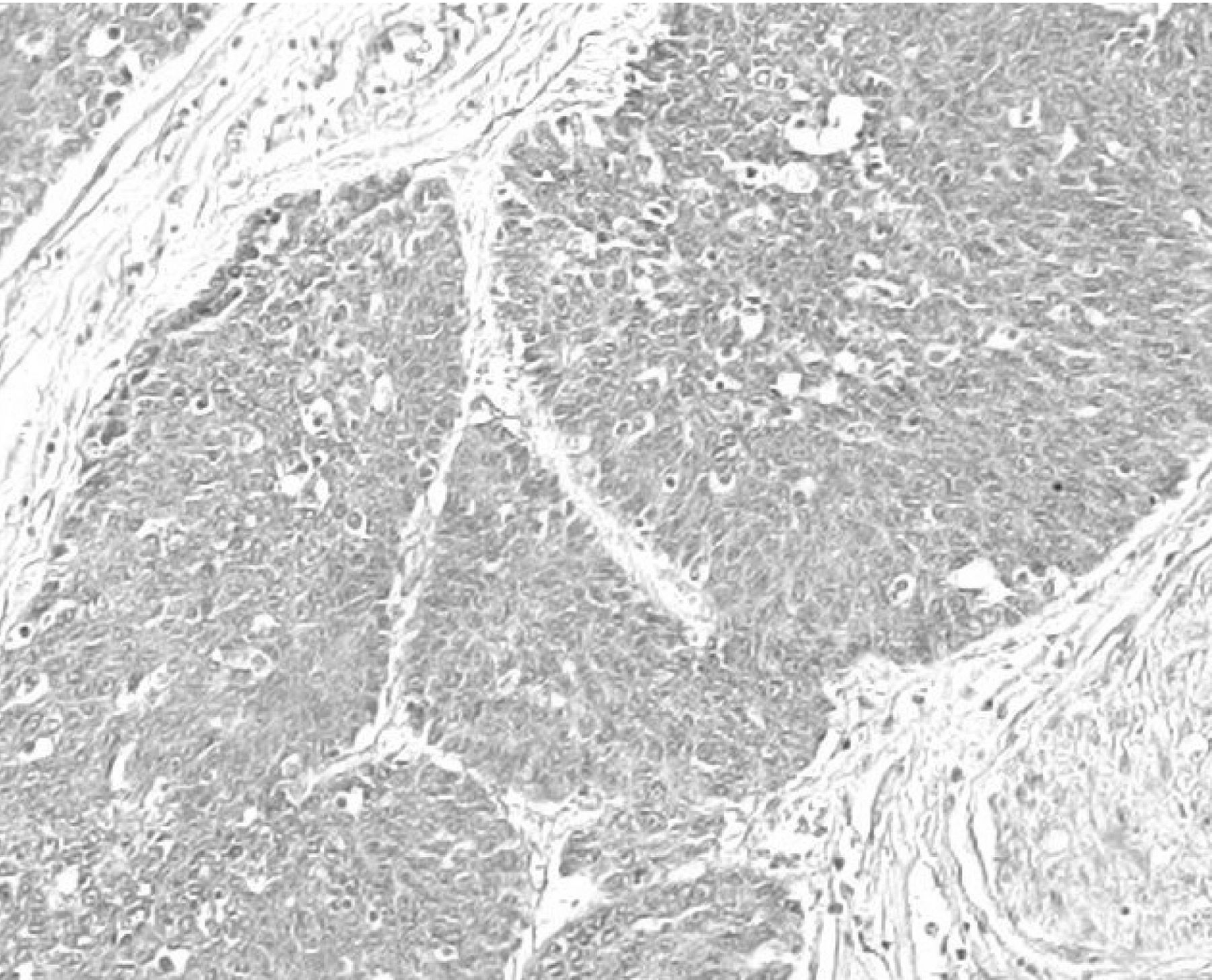
Immunohistochemical detection of tumor interleukin-6 (IL-6) expression. Diffuse cytoplasmic staining of IL-6 protein was detected in tumor cells (magnification, ×100).
Statistical analysis. Comparisons between groups of patients were carried out using the χ2 or Fisher's exact test, as appropriate. Cumulative survival was calculated by the Kaplan–Meier method and survival curves were tested by the log-rank method. Univariate survival analysis was used to estimate the dependence of survival on each variable. Multivariate survival analysis was calculated according to a Cox proportional hazard model in a forward stepwise manner. All statistical analyses were carried out using JMP software (version 8 for Macintosh; SAS Institute Inc., Cary, NC, USA). Significance was defined as a p-value less than 0.05.
Results
Clinicopathological characteristics of study patients. Study patients consisted of 60 patients in the Surgery group and 41 patients in the CRT group, with a median age of 65 (range=44-82) years. A comparison of the clinico-pathological characteristics of patients in the Surgery and CRT groups is shown in Table I. In the pre-treatment parameters, a significant difference between the groups was observed in the tumor location (Cervical esophagus/Upper thoracic esophagus/Middle thoracic esophagus/Lower thoracic esophagus: 2/8/30/20 in the Surgery group vs. 6/8/22/5 in the CRT group), clinical T (T1/T2/T3/T4: 27/12/20/1 in the Surgery group vs. 0/0/16/25 in the CRT group), N, and stage (I/II/III/IV: 26/12/18/4 in the Surgery group vs. 0/2/36/3 in the CRT group). These differences were mostly due to patient selection for induction CRT. All clinical M1 cases were distant lymph node metastases at supraclavicular and celiac regions in both groups. In postoperative pathological parameters, significant differences were observed in pT (T0/T1/T2/T3/T4: 0/33/9/16/2 in the Surgery group vs. 7/1/4/22/7 in the CRT group) and pstage (0/I/II/III/IV: 0/25/16/11/8 in the Surgery group vs. 6/2/12/16/5 in the CRT group). Curability (residual tumor) was not different between the groups. In the CRT group, seven primary tumors were evaluated as grade 3, and 11 and 23 tumors were evaluated as grade 2 and grade 1, respectively.

Tumor interleukin-6 (IL-6) expression in relation to the pathological stage and T factor. Tumor IL-6 expression was almost exclusively detected in advanced tumors of resected specimens of patients in the Surgery group (21.1% of pathological stage III-IV tumors; 27.8% of pathological T3-4 tumors).
Tumor IL-6 expression. Representative tumor IL-6 protein expression is shown in Figure 1. In the resected specimens of patients who underwent esophagectomy without any pre-operative therapies, tumor IL-6 expression was detected in four out of 19 patients (21.1%) with pathological stage III-IV tumors, whereas tumor IL-6 expression was detected in one out of 41 patients with pathological stage I-II tumors. In addition, tumor IL-6 expression was detected in five out of 18 patients (27.8%) with pathological T3-4 tumors, whereas tumor IL-6 expression was not detected in 42 patients with pathological T1-2 tumors (Figure 2).
Survival analysis. The results of univariate survival analysis of overall survival are shown in Table II. Patients with pathological CR (n=7) were excluded from the analysis of the CRT group. In the Surgery group (Table IIA), significant differences in the pre-treatment parameters were observed in cT (T3-4/T1-2), cN (N1/N0), and cstage (III-IV/I-II). No significant differences were observed in the postoperative parameters, excluding pN (N1/N0). Residual tumor (R1-2/R0) and tumor IL-6 expression (positive/negative) were not selected as significant prognostic factors. On the other hand, in the CRT group (Table IIB), no significant differences were observed in any of the pre-treatment parameters, whereas significant differences in post-operative parameters were observed in pT (T3-4/T0-2) and residual tumor (R1-2/R0). Tumor IL-6 expression (positive/negative) was selected as a significant prognostic factor when evaluated by Kaplan–Meier survival analysis (p-value=0.046, Figure 3A). When these three variables (pT, residual tumor, and tumor IL-6) were studied in multivariate analysis, residual tumor and tumor IL-6 were selected as independent prognostic factors (Table III). In addition, to determine the influence of CRT on tumor IL-6 expression, overall survival in patients with clinical T3-4 tumors was calculated according to tumor IL-6 expression, and was compared between the Surgery and CRT groups (Figure 3). Overall survival was not different between patients with and without tumor IL-6 expression in the Surgery group, whereas overall survival in patients with tumor IL-6 expression was significantly shorter than in patients without tumor IL-6 expression in the CRT group.

Kaplan–Meier survival curves according to tumor interleukin-6 (IL-6) expression. A significant difference in overall survival was observed between IL-6-positive and IL-6-negative tumors of resected specimens of patients with cT3-4 tumors who underwent induction chemoradiotherapy followed by esophagectomy (A), whereas no difference was observed in patients with clinical T3-4 tumors who underwent esophagectomy without any preoperative therapy (B).
Univariate analysis of overall survival in the Surgery group.
Relationships between tumor IL-6 and clinicopathological parameters. The results of correlation analysis of tumor IL-6 expression with clinicopathological parameters are shown in Table IV. Patients with pathological CR (n=7) were excluded from the analysis of the CRT group. In the Surgery group (Table IVA), tumor IL-6 expression was significantly correlated with each of cT, pT, pstage, and residual tumor, whereas there was no significant correlation between tumor IL-6 and any of the clinicopathological factors in the CRT group (Table IVB).
Univariate analysis of overall survival in the Chemoradiotherapy (CRT) group.
Multivariate analysis of overall survival in the chemoradiotherapy group.
Discussion
Elevated serum IL-6 levels have been associated with disease progression and poor prognosis in patients with esophageal carcinoma (2-4). However, there have been few reports on tumor IL-6 expression in relation to tumor progression and survival. Our previous study has demonstrated a correlation between elevated serum IL-6 levels after induction CRT, and not before CRT, and resistance to CRT and poor prognosis in patients with advanced ESCC who underwent induction CRT followed by surgery (7). That study also confirmed that serum IL-6 levels correlated with tumor size in resected specimens after CRT; tumor IL-6 expression was detected by immunohistochemistry within the tumor tissues. These findings suggested that serum IL-6 may reflect tumor IL-6 expression and that the tumor microenvironment associated with poor patient outcomes. The present study was conducted to test this hypothesis, and demonstrated that tumor IL-6 expression, after CRT, predicts poor patient outcome.
Relationship between tumor interleukin-6 (IL-6) expression and clinicopathological factors in the surgery group.
It has been shown that cancer progression involves the expression of inflammatory cytokines, including IL-6, within the tumor microenvironment, and such cancer-related inflammation mediates tumor invasion and metastasis, angiogenesis, and immunosuppression, thereby promoting cancer progression (10, 11). This is in agreement with our finding that tumor IL-6 expression in the resected specimens of patients who underwent surgery alone was detected in advanced tumors (high pT and pstage). In addition, when overall survival was compared in patients with cT3-4 tumors, tumor IL-6 expression was shown to be a significant indicator of poor prognosis in patients who underwent CRT followed by surgery, whereas it was not in patients who underwent surgery alone. This suggests that CRT may induce qualitative alterations in tumor cells or within the tumor microenvironment associated with CRT resistance and poor survival. Two possibilities may explain the prognostic significance of tumor IL-6 expression in the CRT group; one is that IL-6 may act originally as a resistance factor against CRT, and IL-6-negative tumor cells sensitive to CRT may be eliminated during CRT. In fact, IL-6 has been reported to act as a resistance factor against irradiation in several cancer types (12, 13). However, it is difficult to explain CRT resistance by IL-6 alone because 24 out of 34 residual tumors after CRT were negative for IL-6. Another explanation is that IL-6 may be induced or overexpressed by CRT, and act as a CRT resistance factor or as a predictor of poor prognosis. In this regard, nuclear factor-kappa B (NF-κB) status has been associated with the CRT response and prognosis in patients with esophageal adenocarcinoma who underwent pre-operative CRT (14-16). NF-κB is a transcription factor that plays a critical role in cancer progression, and IL-6 is a downstream molecule regulated by NF-κB (17). Izzo et al. demonstrated that the CRT-induced increase in activated NF-κB was more significantly correlated with a poor response and prognosis than the pre-treatment presence of activated NF-κB using immunohistochemical staining for activated NF-κB in pre-treatment biopsy specimens and post-treatment surgical specimens (14, 15). In addition, it has been reported that IL-1β and IL-8 protein expressions in tumor tissue homogenates between pre- and post-CRT were significantly down-regulated in responders, whereas no differences were detected in non-responders (16). These findings strongly suggest that CRT-induced alterations in the NF-κB status and the expression of pro-inflammatory cytokines regulated by NF-κB may determine CRT resistance and poor outcomes. These findings also seem to be in line with our findings that there is a prognostic difference in tumor IL-6 expression according to the presence or absence of CRT.
Relationship between tumor IL-6 expression and clinicopathological factors in the chemoradiotherapy group.
In conclusion, the present study demonstrated for the first time that tumor IL-6 expression increases in proportion with primary tumor progression, and that CRT-induced tumor IL-6 expression has an independent prognostic value. However, the present study did not perform a comparison between pre- and post-CRT specimens. In addition, there was a selection bias between patients with and without pre-operative CRT despite the identical clinical T3-4 tumors. In the near future, the clinical significance of tumor IL-6 expression will be elucidated in a prospective study by using pre- and post-CRT specimens in combination with assessment of NF-κB and other pro-inflammatory cytokines.
- Received April 16, 2013.
- Revision received May 2, 2013.
- Accepted May 9, 2013.
- Copyright© 2013 International Institute of Anticancer Research (Dr. John G. Delinassios), All rights reserved
References
In this issue


Jump to section
Related Articles
Cited By...
- No citing articles found.