


Abstract
Background/Aim: We have previously reported that alternate-day S-1 had comparable effects and milder adverse events than the respective consecutive-day regimen in head and neck cancer (HNC) patients. The aim of this study was to investigate the anticancer effects of both regimens and underlying mechanisms in vitro. Materials and Methods: Two head and neck squamous cell carcinoma (HNSCC) cell lines were treated with 5-FU given on an alternate-day or consecutive-day schedule. The relative inhibition (RI) of tumor growth was calculated. Cell cycle distributions and cyclin expression following 5-FU treatment were analyzed. Results: The RI of both regimens was almost identical. The percentage of cells in S phase was significantly increased in the alternate-day group compared to the consecutive-day group (p<0.001). Conclusion: The cytotoxic effect of alternate-day was equivalent to that of consecutive-day. S-phase arrest was more prominently observed with the alternate-day regimen, which may help maintain 5-FU sensitivity in head and neck cancer cells.
Head and neck squamous cell carcinoma (HNSCC) is still lethal because of the high percentage of recurrence and metastasis (1-4). S-1 is a novel oral anticancer agent consisting of tegafur, 5-chloro-2,4-dihydroxypyridine, and potassium oxonate at a molar ratio of 1:0.4:1, based on biochemical modulation of 5-fluorouracil (5-FU) (1, 5). A phase III study [Adjuvant Chemotherapy with S-1 after Curative Treatment in Patients with Head Neck Cancer (ACTS-HNC) trial] investigated whether adjuvant therapy with S-1 could improve the prognosis in head and neck cancer (6). S-1 was administered daily for 2 weeks, followed by a 1-week rest period. The study showed that adjuvant chemotherapy with S-1 resulted in significantly better overall survival (OS) in patients with locally advanced HNSCC compared to the control arm. However, the S-1 arm was less well-tolerated and the 1-year completion rate of S-1 was only 43.4%. Therefore, treatment completion and tolerance remain unresolved problems. Sakuma et al. reported that S-1 given on an alternate-day schedule can reduce the incidence of adverse effects without compromising the therapeutic effects of S-1 (7). The alternate-day regimen was equivalent to or better than the consecutive-day regimen in patients with other regimens for metastatic pancreatic cancer in a clinical trial (8). Likewise, we conducted a retrospective study of an alternate-day S-1 regimen as adjuvant therapy after surgery and chemoradiotherapy for advanced HNSCC. In that study, we observed that alternate-day S-1 administration caused fewer adverse effects and was tolerable for patients with advanced HNSCC (9). The completion rate was 80.7% and the occurrence of adverse events tended to be less than that reported in the ACTS-HNC trial. Three-year OS and disease-free survival rates were 74.8% and 57.3%, respectively. Other groups also reported that alternate-day S-1 treatment was associated with milder adverse events without compromising therapeutic effectiveness in clinical or pre-clinical studies in gastric cancer or pancreatic cancer (7, 10). Thus, alternate-day S-1 regimen may be an effective adjuvant treatment with milder side effects than the original consecutive-day regimen. Here, we investigated anticancer effects and underlying mechanisms of 5-FU given on an alternate-day schedule compared with the consecutive-day schedule in head and neck cancer cells in vitro.

Treatment schedules of 5-fluorouracil were planned to estimate tumor growth inhibition in vitro. For consecutive-day treatment, 5-fluorouracil was added on days 1-3 and culture medium was added on days 4-6. For alternate-day treatment, cell cultures were exposed to 5-fluorouracil or culture medium every other day (5-fluorouracil was added on days 1, 3, and 5). The time point of the equivalent total dose is day 6.
Materials and Methods
Cell lines and reagents. The HSC-3-M3 cell line (11-13), derived from a patient with oral squamous cell carcinoma, was obtained from the Japanese Collection of Research Bioresources (Osaka, Japan). HSC-3-M3 cells were cultured in Eagle's minimum essential medium supplemented with 10% fetal bovine serum (FBS), 100 units/ml penicillin, and 100 units/ml streptomycin in a humidified incubator with 5% CO2 at 37°C. The BICR6 cell line (14), derived from a patient with hypopharyngeal cancer, was obtained from the European Collection of Cell Cultures (United Kingdom). BICR6 cells were cultured in Dulbecco modified Eagle's medium supplemented with 10% FBS, 0.4 μg/ml hydrocortisone, 100 units/ml penicillin, and 100 μg/ml streptomycin in a humidified incubator with 8% CO2 at 37°C.
5-FU and dimethyl sulfoxide (DMSO) were purchased from Wako (Tokyo, Japan); stock solutions of 5-FU were made in DMSO and stored at −20°C until use. The final concentration of DMSO was <0.1% in the culture medium and the same concentration of DMSO was present in the control group.
Cell doubling time. HSC-3-M3 and BICR6 cells were seeded onto flat-bottom 10-cm dishes (NIPPON Genetics, Tokyo, Japan) at a density of 20,000 cells/dish. Doubling time was calculated using the following formula=[T×loge2]/[ln(Y/X)], where T is time in any unit, X is starting number of cells, and Y is final number of cells after trypsinization. Proliferation of HSC-3-M3 and BICR6 cells was analyzed by counting the total cell number at 0, 24, 48, and 72 h (15).
Proliferation assay. HSC-3-M3 and BICR6 cells were seeded onto 24-well flat-bottom microdilution plates (Sumitomo Bakelite, Tokyo, Japan) at a density of 1,500 cells/well. Cell suspensions were then transferred to 8 wells each for treatment and control groups. Cell cultures were statically washed three times with culture solution once daily. For consecutive-day treatment, 5-FU was added on days 1-3 and culture medium was added on days 4-6. For alternate-day treatment, cell cultures were exposed to 5-FU or culture medium every other day (5-FU was added on days 1, 3, and 5). The control group was exposed to culture medium only. The number of viable cells was determined by addition of WST-8 (Dojindo Laboratories, Kumamoto, Japan) according to the manufacturer's instructions. Data were collected from three independent experiments. Time courses are shown in Figure 1. Half-maximal inhibitory concentration (IC50) values, i.e., drug concentrations at 50% cell growth inhibition compared with control cell growth, were used in all subsequent experiments.
Inhibition analysis of tumor growth in vitro. HSC-3-M3 and BICR6 cells were seeded onto 24-well flat-bottom microdilution plates (Sumitomo Bakelite) at a density of 1,500 cells/well. Cell suspensions were then transferred to 8 wells each for treatment and control groups. Cell cultures were statically washed three times with culture solution once daily. Time courses were same as described above for the proliferation assay. The optical density in the treatment group was divided by that in the control group to calculate the tumor growth ratio relative to control (T/C). The relative inhibition (RI) rate of tumor growth was calculated by the following equation: RI (%)=(1−T/C)×100.

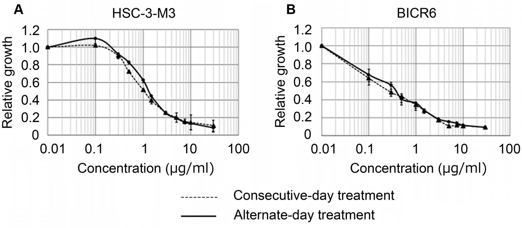
In vitro responses of HSC-3-M3 and BICR6 cells to effects of various concentrations of 5-fluorouracil. Cell viability was assessed using the WST-8 assay.
Cell cycle analysis by flow cytometry. Cell cycle analysis is one of the most applied protocols in flow cytometry and is most commonly used to measure frequencies of cells that are either in G0/G1, G2/M, or S phase of the cell cycle. To determine the percentage of cells in various phases of the cell cycle, exponentially proliferating cells were treated with 5-FU. Treated cells and untreated controls were then analyzed for nuclear DNA after propidium iodide staining using the Cycletest™ Plus DNA Reagent kit (Becton Dickinson, San Jose, CA, USA) according to the manufacturer's instructions. This kit provides a set of reagents for isolating and staining cell nuclei from surplus cell suspensions. Flow cytometric analysis of differentially stained normal and tumor cells was then used to identify abnormal DNA stemlines and estimate the DNA index (DI) and cell-cycle phase distributions of these stemlines. Flow cytometric analysis was performed using the FACSCalibur™ flow cytometer (Becton Dickinson).
Western blot analysis. Treated cells and untreated controls were washed twice with ice-cold phosphate-buffered saline and then harvested by scraping in Lysis buffer (Cat. No. C3228; Merck, Kenilworth, NJ, USA). After homogenization and centrifugation for 30 min at 13,000 × g, the supernatants (20 μg of protein) were subjected to SDS-PAGE, followed by transfer to polyvinylidene difluoride membranes (Cat. No. IPVH00010; Merck). Immunoblot analysis was performed on day 7 of treatment using the following primary antibodies: cyclin A (1:200 dilution; ab38), cyclin D (1:20,000 dilution; ab134175), cyclin E (1:1,000 dilution; ab133266), and β-actin (1:1,000 dilution; ab8226), which were obtained from Abcam (Cambridge, UK). After washing, cells were incubated with horseradish peroxidase-conjugated anti-rabbit IgG or anti-mouse IgG secondary antibody (Jackson ImmunoResearch Laboratories, West Grove, PA, USA). Images were obtained using Fujifilm LAS-4000 mini (Fujifilm, Tokyo, Japan). Quantification of band density was obtained by ImageJ software (Version 1.5).
Statistical analysis. All data were obtained from triplicate experiments and are expressed as mean±standard deviation from triplicate experiments. Student's t-tests were employed for analyzing data. All statistical analyses were performed with EZR (13); p-values <0.05 were considered statistically significant.
Results
Cell doubling time. Doubling times of tumor cell lines were 24.4 h for HSC-3-M3 and 30.6 h for BICR6.
Determination of IC50 in HSC-3-M3 and BICR6 cells. Antiproliferative effects of 5-FU in HSC-3-M3 and BICR6 cells were determined by the WST assay. The drug inhibited cell growth in a dose-dependent manner and both treatment schedules inhibited cell growth to a comparable degree (Figure 2). IC50 of HSC-3-M3 and BICR6 were 1.5 μg/ml and 0.4 μg/ml, respectively, in alternative-day treatment.
Inhibition of cell proliferation in vitro. In this study, the experiments were conducted at the same concentration to clarify the differences in growth inhibitory effects when only the administration schedule was changed. Thus, we performed the following experiment at IC50 concentration in alternative-day treatment. The RI of consecutive-day treatment in BICR6 and HSC-3-M3 cell lines was 62.6±0.6% and 47.2±5.3%, respectively, while the RI of alternate-day treatment in BICR6 and HSC-3-M3 cell lines was 61.6±0.7% and 46.5±1.5%, respectively. The difference in the RI between consecutive-day treatment and alternative-day treatment was not statistically significant (Table I). Saga et al. reported that the drug-free interval in the intermittent infusion of 5-FU had to be shorter than the doubling time of targeted tumor cells to obtain a similar killing effect as continuous administration in vitro (14). As shown above, the two cell lines tested showed doubling times of >24 h; thus, we speculated that alternative-day 5-FU treatment achieved a similar killing effect as consecutive-day treatment in the two cell lines.
Antitumor effect of 5-fluorouracil (5-FU) on the HSC-3-M3 and BICR6 cell lines.
Alternate-day treatment with 5-FU prolonged the S phase in HSC-3-M3 and BICR6 cells compared to consecutive-day treatment and control. 5-FU only targets cycling cells and modulates cancer cell cycle status, and 24-h exposure to 5-FU is associated with an accumulation of cells in S phase (1). The population of HSC-3-M3 cells in S phase on day 4 of treatment was 25.9±2.2% in the control group, 75.2±3.8% in the consecutive-day treatment group, and 55.4±5.4% in the alternate-day treatment group. The population of BICR6 cells in S phase on day 4 of treatment was 32.8±2.6% in the control group, 68.7±5.4% in the consecutive-day treatment group, and 58.9±5.9% in the alternate-day treatment group (Figure 3A). The population of HSC-3-M3 cells in S phase on day 7 of treatment was 15.1±2.5% in the control group, 22.7±4.5% in the consecutive-day treatment group, and 54.3±6.8% in the alternate-day treatment group. The population of BICR6 cells in S phase on day 7 of treatment was 20.0±3.5% in the control group, 38.3±3.7% in the consecutive-day treatment group, and 57.1±4.8% in the alternate-day treatment group (Figure 3B). In consecutive-day treatment, the percentage of cells in S phase on day 7 of treatment was significantly decreased compared to that on day 4 of treatment in HSC-3-M3 and BICR6 cells (p<0.05) (Figure 3C). In alternate-day treatment, the percentage of cells in S phase on day 7 of treatment was almost the same as that on day 4 in HSC-3-M3 and BICR6 cells (n.s.: not significant) (Figure 3D). There was a significantly increased percentage of cells in S phase on day 7 of treatment with the alternate-day regimen compared with control or consecutive-day regimen in HSC-3-M3 and BICR6 cells (p<0.05) (Figure 3D).
Cyclin expression. The aforementioned effect of increased percentages of S phase cells with alternate-day treatment may be explained by S phase arrest. Therefore, cyclin A/D/E expression in HSC-3-M3 cell lines was examined by western blotting analysis on day 7 of treatment from triplicate experiments (Figure 4). In the alternate-day regimen group, cyclin A and E expression levels were increased and cyclin D expression were decreased compared to that of the consecutive-day regimen. The presented image bands are representative of 3 independent experiments in Figure 4A. All the experiments showed the same trend, however differences were not statistically significant (Figure 4B). These results of western blot analysis are consistent with the cyclin expression pattern reported in S phase arrest (18, 19).
Discussion
We showed that the cytotoxic effect of alternate-day 5-FU treatment was not significantly different from that of consecutive-day 5-FU treatment in HSC-3-M3 and BICR6 cells. However, the percentage of S-phase cells on day 7 of treatment was significantly increased in the alternate-day regiment compared to that of the consecutive-day regimen, in both cell lines. In the alternate-day regimen, cyclin A and E expression was increased and cyclin D expression was decreased compared with that of the consecutive-day regimen. Shirasaka et al. examined the alternate-day S-1 regimen and the relation between cell cycle length and antitumor activity of 5-FU (20). They stated that the theoretical basis by which alternate-day S-1 administration exerts its effect is as follows. Normal cells and tumor cells have different cell cycles in vivo. The cell cycle of normal cells is only 0.5-1.5 days, and the length of S phase, in which 5-FU was activated, was 9-14 h (21). In contrast, the cell cycle of tumor cells is 5-7 days and S phase lasted for >17-60 h. Therefore, drug withdrawal for 24 h allows proliferation of normal cells that do not encounter 5-FU during their shorter S phase, while cancer cells are still exposed to 5-FU during their longer lasting S phase (20). In a study in which ovarian cancer cells were given intermittent dosing with 5-FU withdrawal time and consecutive administration, Saga et al. reported that the therapeutic effect of 5-FU is impaired when the intermittent administration withdrawal time is longer than the doubling time of cancer cells (17). The doubling times of HSC-3-M3 and BICR6 used in the present study were both > 24 h. The resting time of 5-FU was set to 24 h. Our experimental result was consistent with the report by Saga et al. In flow cytometry, the proportion of cells in S phase on day 7 of treatment increased with alternate-day administration compared to consecutive-day administration. 5-FU only targets cycling cells and modulates cancer cell cycle status via one of the following three modes: i) loss or accumulation of S-phase cells; ii) G2/M block; and iii) G1/S arrest (22). In our case, the cyclin expression pattern suggested that the alternate-day 5-FU regimen caused S phase arrest (18, 19). Because 5-FU acts on S-phase cells, S-phase arrest may increase susceptibility to the drug. In order to further improve the effectiveness and tolerability of alternate-day 5-FU administration in head and neck cancer, further examination such as dose setting is warranted.

Cell cycle distributions were analyzed by flow cytometry. DNA distribution histogram using propidium iodide (PI) labeling (x-axis) and total number of cells in each channel (y-axis) in control, consecutive-day 5-fluorouracil (consecutive) treatment, or alternate-day 5-fluorouracil (alternate) treatment cells. Cell cycle distributions were analyzed by flow cytometry (A, B). In consecutive-day treatment, the percentage of cells in S phase on day 7 was significantly decreased (p<0.05) compared with that on day 4 (C). By contrast, in alternate-day treatment, the percentage of cells in S phase on day 7 was almost the same as that on day 4 (D). Thus, the percentage of S phase cells on day 7 was higher (p<0.05) in the alternate-day regimen compared with the consecutive-day regimen or control (E).

Protein levels of cyclins A, D, and E in HSC-3-M3 cell lines were assessed by western blot analysis. Expression levels of cyclins A/E were increased in the alternate-day regimen compared to that in the consecutive-day regimen. Conversely, cyclin D expression was decreased in the alternate-day regimen compared to that in the consecutive-day regimen. Protein levels were quantified using β-actin as an internal control and were normalized to the control group (A). Data are presented as mean relative fold change compared to control±standard deviation and p-values (B).
Acknowledgements
We thank Dr. Ryuichiro Araki (Saitama Medical University International Medical Center) for assistance with statistical analysis. We also thank Ms. Noriko Akiyama (Saitama Medical University International Medical Center) and Mr. Takashi Yamaga (Kyorin University) for technical assistance.
This study was supported by a grant from Setsuro Fujii Memorial, The Osaka Foundation for Promotion of Fundamental Medical Research. The sponsors had no direct impact on study design or conduct; data collection, management, analysis, or interpretation; or preparation, review, or approval of the manuscript.
Footnotes
Authors' Contributions
YM conceived and carried out the experiments with the collaboration of YK, HS, RT, TK, MH, and YM. YM wrote the first draft. HS, YM, YK, and NK reviewed and revised the article.
- Received August 15, 2019.
- Revision received September 17, 2019.
- Accepted September 27, 2019.
- Copyright© 2019, International Institute of Anticancer Research (Dr. George J. Delinasios), All rights reserved


