


Abstract
Background: Due to use of Tween-80 as an enhancer of solubility, the current clinical formulation of cabazitaxel (CBT) (Jevtana®) causes hypersensitivity, neurotoxicity and other severe side-effects. To reduce these vehicle-related effects, a suitable nanocarrier is needed. Materials and Methods: Human serum albumin (HSA) was used to encapsulate CBT by a simple self-assembly method. Physicochemical properties of HSA–CBT nanoparticles were characterized. In vitro release property and cytotoxicity were also determined. In vivo imaging system was used to study nanocarrier distribution in vivo. The safety profile was assessed by hemolysis and acute-toxicity study. Finally, the antitumor efficacy in vivo was investigated in tumor-bearing mice. Results: The average size of HSA-CBT nanoparticles was about 240 nm and the encapsulation efficiency reached 97%. The hemolysis and acute-toxicity experiments confirmed biocompatibility of HSA-CBT nanoparticles. Conclusion: HSA nanoparticles are a safe and effective drug delivery system for hydrophobic anticancer drugs such as CBT.
Taxanes, such as paclitaxel and docetaxel, are widely used in the clinic for treatment of a broad spectrum of cancer types. Cabazitaxel (CBT), a semi-synthetic taxane derivative, is increasingly used in cancer therapy because of its superior pharmacological properties and low drug resistance (1-3). Since CBT is lipophilic and insoluble in water, polysorbate 80 (Tween-80) is used in its clinical formulation, which causes several major side-effects, such as hypersensitivity reactions (4).
To increase solubility and reduce side-effects, several nanoparticle-based delivery systems were developed for CBT, including liposomes, lipid emulsion and polymeric micelles (5, 6). Use of an HSA-based nanoparticle has become an excellent strategy for anticancer drug delivery (7-10). Abraxane is the first successful example of a HSA-based drug-delivery vehicle (11). HSA-bound anticancer drug complexes are capable of using the natural albumin transport pathways. Glycoprotein 60 and caveolin-1-mediated transcytosis can increase the intratumoral accumulation of such complexes. Furthermore, deep penetration into tumor can be achieved through association with tumor-derived secreted protein, acidic and rich in cysteine (SPARC) protein, and overexpression of SPARC is observed in multiple tumor types such as those of the breast, prostate, colorectal, liver and lung (12-20).
To develop a solvent-free delivery system, HSA nanoparticles were used for CBT in this study. The physicochemical characteristics of HSA–CBT nanoparticles (NPs) were measured. Its in vitro drug-releasing behavior and cytotoxicity were characterized in cancer cell lines. Moreover, for future clinical translation, the pharmacokinetics, drug distribution, antitumor efficacy and tolerability profile of the nanoparticles were examined in vivo.
Materials and Methods
Materials. CBT was purchased from Yew Biotechnology Co. (Jiangsu, China). Penicillin-streptomycin, RPMI-1640, fetal bovine serum (FBS), 0.25% (w/v) trypsin, and 0.03% (w/v) EDTA solution were purchased from Hyclone (USA). 1,1-dioctadecyl-3,3,3,3-tetramethylindotricarbocyanine iodide (DiR iodide) (D12731) probe was purchased from Life Technologies (Shanghai, China). HSA and 3-(4,5-dimethyl-thiazol-2-yl)-2,5-diphenyltetrazolium bromide (MTT) were purchased from Sigma-Aldrich (Shanghai, China).
Nanoparticle preparation. To prepare the HSA–CBT-NPs, HSA (10 mg, 20 mg, 30 mg corresponding to the formulation of H10-1, H10-2, H10-3, respectively) was dissolved in 1.8 ml deionized water and 1 mg CBT was dissolved in 0.1 ml ethanol. The CBT solution was added to the HSA solution under stirring and incubated at room temperature for 15 min. The ethanol in the mixed solution was removed by vacuum rotary evaporation at 37°C.
Size, zeta potential and morphological determination. The characteristics of HSA–CBT-NPs including volume-average diameter and zeta potential (ξ) were measured by a Malvern Zetasizer (Nano-ZS, Malvern, Worcestershire, UK) at room temperature. Their morphology was observed through a transmission electron microscope (JEM-200C; JEOL, Tokyo, Japan) at an acceleration voltage of 200 kV.
Drug-loading capacity and encapsulation-efficiency assays. To determine the concentration of free CBT, one aliquot of the HSA–CBT-NPs was ultrafiltered on a Centricon filter (4,000 × g, 5 min) and 200 μl of the filtrate was mixed with 800 μl acetonitrile, and vortexed for 3 min. An aliquot of the same HSA–CBT-NPs (200 μl) was mixed with 800 μl acetonitrile and vortexed for 3 min without other process to determine the concentration of total CBT. CBT concentration was then measured by high performance liquid chromatography (HPLC). A Hypersil ODS-2 C18 reverse phase column (5-μm particle size, 250 mm×4.6 mm) (Dalian Elite Analytical Instruments Co., Ltd., Dalian, China) was used in the analysis and the mobile phase used consisted of 60% (v/v) acetonitrile and 40% deionized water as an isocratic system. Flow rate was 1.0 ml/min and the effluent was monitored at 230 nm. Encapsulation, drug-loading capacity and encapsulation efficiency were calculated as follows:
 (Eq. 1)
 (Eq. 2)
 (Eq. 3)
In vitro release assay. To investigate the release properties of HSA–CBT-NPs, in vitro release was carried out by dialysis method in phosphate-buffered saline (PBS) (0.2% Tween-80, pH 7.4).
HSA–CBT-NPs (1.0 ml, 1 mg CBT equivalent) were diluted with 9.0 ml PBS and 1.0 ml solution was taken to determine the initial concentration. The remaining 9.0 ml solution was placed in dialysis tubing (MWCO=14 kDa) and immersed into 50 ml PBS. The release condition used was shaking at 100 rpm at 37°C for 2 days. A volume of 1.0 ml release medium was sampled at designated times (1, 2, 4, 8, 12, 24, 36 and 48 h) followed by immediately replacement with an equal volume of fresh PBS. The concentration of CBT in samples was determined by HPLC. A formulation used in Jevtana®, containing ethanol and Tween-80, was used as a control.
In vitro cytotoxicity. HCT116 Human colonic cancer cells and A549 human lung adenocarcinoma cells were cultured in RPMI-1640 supplemented with 10% (v/v) FBS and 1% (v/v) antibiotics at 37°C, in a humidified atmosphere with 5% CO2. In order to evaluate the cytotoxicity of HSA–CBT-NPs, inhibition of cell growth was determined by MTT assay. HCT116 and A549 cells were seeded into 96-well plates at 8×10³ cells/well and incubated at 37°C, in a humidified atmosphere with 5% CO2 for 24 h. The culture medium was then changed to that containing different concentrations (0.2 to 100 μg/ml) of HSA–CBT-NPs suspended in RPMI-1640. RPMI-1640 medium was used as a blank control and the formulation in Jevtana® was used as control group. After incubation for 48 h, cell viability was determined by the MTT assay. Briefly, 30 μl of MTT solution (5 mg/ml) were added to each well. After another 4 h incubation, the medium was replaced with dimethyl sulfoxide (100 μl/well) to dissolve the formazan product. The optical density of the solution was determined by a microplate reader (SpectraMax M4; Molecular Devices, Silicon Valley, CA, USA) at 492 nm.
In vitro hemolysis. Blood from Sprague-Dawley rats was used for evaluating the hemolysis of HSA–CBT-NPs, while the CBT formulation used in Jevtana® was used to compare the biocompatibility. To obtain red blood cells (RBCs), fresh blood was centrifuged at 1,500 × g for 10 min to remove the plasma. Then the RBCs were washed three times with normal saline and collected by centrifugation, and diluted to a density of 2% (v/v). Different concentrations of HSA–CBT-NPs and Jevtana® were added to the RBC suspension (1:1, v/v), normal saline was added to the RBCs as a negative control, while distilled water was added as a positive control. After incubation for 1.5 h at 37°C, the RBC suspension was centrifuged at 1,500 × g for 10 min and 150 μl supernatant was added to 96-well plates. A microplate reader (SpectraMax M4; Molecular Devices) was used to measure the absorbance (Abs) at 545 mM and the hemolysis rate was calculated as follows:
 (Eq. 4)
In vivo pharmacokinetic studies. To investigate the pharmacokinetics of HSA–CBT-NPs, 10 male Sprague-Dawley rats (mean±SD=250±20 g) were divided into groups at random (n=5) and then intravenously injected with HSA–CBT-NPs, while CBT injection was used as control. Rats in each group were given a single 8 mg/kg dose (CBT equivalent), and blood samples were collected at different time points (5, 15, 30 min and 1, 2, 4, 8, 24 h). All samples were collected into heparinized tubes and then centrifuged at 4,000 × g for 10 min to obtain plasma; 200 μl of plasma was transferred into 2 ml centrifuge tubes and then 50 μl of the internal standard diazepam (National Institutes for Food and Drug Control, Beijing, China) (10 μg/ml acetonitrile) was added. Next, the samples were centrifuged at 13,000 × g for 10 min after 350 μl of acetonitrile was added with vigorous vortex mixing. CBT concentration in the supernatant was measured by HPLC.
In vivo distribution study. To observe HSA–CBT-NPs distribution after intravenous administration by in vivo imaging system (IVIS), DiR-loaded nanoparticles were prepared as described in Nanoparticle preparation. Three nude mice bearing A549 tumor (approximately 1,000 mm3) were injected with 0.2 ml of DiR-loaded nanoparticles (100 μg/ml) via tail vein and fluorescent images were obtained at 4 h using a Clairvivo OPT instrument (Shimazu Corporation, Kyoto, Japan) with a 735 nm single laser and 5 s exposure time after mice were anesthetized by 10% hydrated chloral solution. Then the mice were euthanized and major organs were excised (heart, liver, spleen, lung, kidney and tumor), and fluorescence signal intensity in different tissues was measured.
Mean±SD size and zeta potential of HSA–CBT nanoparticles (n=3).

Particle size distribution and transmission electron microscopy images of HSA–CBT nanoparticles. a-c: Particle size distribution of H10-1, H10-2 and H10-3, respectively; d and e:TEM images of H10-1 and H10-3. Scale bar is 0.2 μm.
In vivo antitumor efficacy. To evaluate the antitumor efficacy of HSA–CBT-NPs, in vivo tumor growth-inhibition was evaluated. Twenty-one BALB/c nude mice bearing HCT116 tumor were randomly divided into three groups (n=7) and the formulations, or normal saline and CBT injection as controls, were given intravenously via the tail vein every 3 days four times. Tumor volume measured as:
 (Eq. 5)
and body weight were measured regularly as an indicator of antitumor efficacy and acute toxicity, respectively.
(Eq. 5)
and body weight were measured regularly as an indicator of antitumor efficacy and acute toxicity, respectively.
Acute-toxicity experiment. To study the formulation's toxicity, HSA–CBT-NPs was given as a single injection to five healthy male mice at a concentration of 10 mg/kg (CBT equivalent) via the tail vein. Normal saline was used as negative control, while CBT injection was used as positive control, respectively. Body weight changes of the mice were measured for a week then their blood samples were taken to determine the routine blood and biochemical parameters.

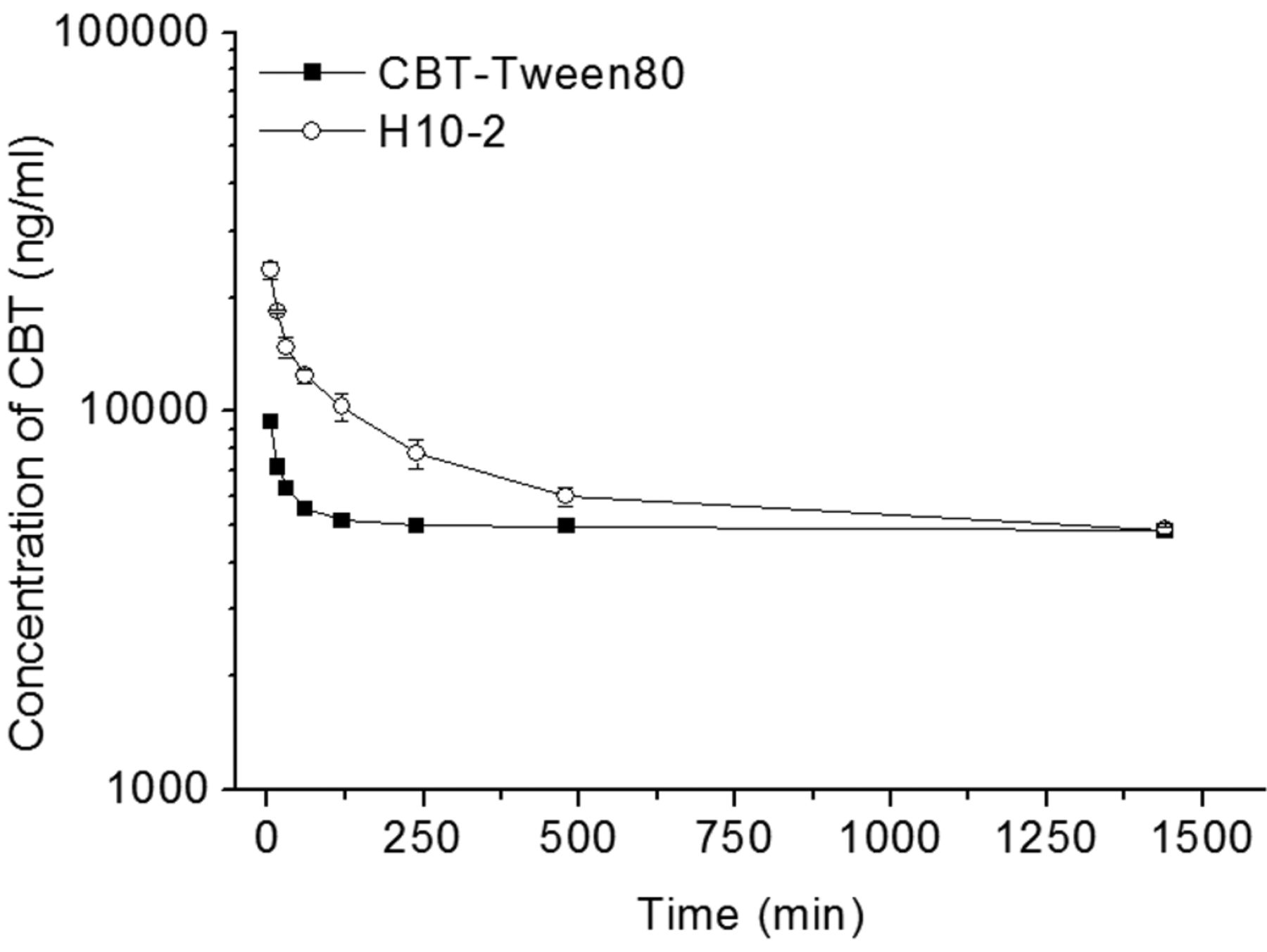
Cumulative release curve of different formulations of HSA–CBT nanoparticles. Nanoparticles were placed in phosphate-buffered saline (pH 7.4) medium containing 0.2% Tween-80 at 37°C.

In vitro cytotoxicity of different formulations of HSA–CBT nanoparticles against human HCT116 (a) and A549 (b) cancer cells (data are mean±SD, n=3).
Results
Characterization of nanoparticles. HSA–CBT-NPs were prepared from HSA and CBT in different proportions and their particle size and zeta potential are summarized in Table I. The size of HSA-NPs and zeta potential depended on the ratio of HSA to CBT. The drug-loading capacity and encapsulation efficiency of HSA–CBT-NPs (H10-2) were determined to be approximately 4.65% and 97.7%, respectively. The size distribution and TEM images of HSA–CBT-NPs are shown in Figure 1 and demonstrate the good dispersibility and uniform size of the nanoparticles.
In vitro release behavior of HSA–CBT-NPs. The release properties of HSA–CBT-NPs compared with CBT injection and the contrast among different proportions are shown in Figure 2. The release properties of HSA–CBT-NPs were similar to those of CBT injection and nearly 100% of the loading drug was released.

In vitro hemolysis. a: Image of hemolysis resulting from HSA–CBT nanoparticles and Jevtana® after centrifuging. b: The percentage of hemolysis induced by HSA–CBT-NPs and Jevtana® (data are mean±SD, n=3).
Cell-viability assay. HCT116 and A549 cell lines were used for the MTT assay. The relationship between drug concentration and cell viability are shown in Figure 3. All the formulations had cytotoxic activity and the half-maximal inhibitory concentrations (IC50) of HSA–CBT-NPs were 25 μg/ml for HCT116 cells and 90 μg/ml for A549 cells.
In vitro hemolysis. Tween-80 and ethanol, commonly used in CBT injection, can cause hemolysis. However, HSA–CBT-NPs should overcome this problem. To confirm this, an in vitro hemolysis experiment was carried out. The results in Figure 4 clearly show that little hemolysis was observed using HSA–CBT-NPs until the concentration reached 500 μg/ml. This result confirmed that HSA–CBT-NPs are safer and have better biocompatibility than CBT injection.
Pharmacokinetics of HSA-NPs. Compared with Jevtana®, HSA–CBT-NPs maintain a higher plasma concentration of CBT until 8 h (Figure 5) and the initial plasma concentration of CBT released from HSA–CBT-NPs was much higher than that of Jevtana®. The values of the area under the curve (AUC) for HSA–CBT-NPs were significantly higher, based on the slow-release function of NPs which extends the circulation time of CBT.
In vivo distribution study. The biodistribution of DiR-loaded HSA nanoparticles clearly illustrates their tumor-targeting ability (Figure 6). Four hours after injection, significant signals in the tumor site were observed. HSA nanoparticles were shown to be an efficient delivery system.

Pharmacokinetics of HSA–CBT nanoparticles and Jevtana® (n=5).
In vivo antitumor efficacy. BALB/c nude mice bearing HCT116 tumor were used to verify the antitumor efficacy and the results are shown in Figure 7. Similar antitumor efficacy between HSA-CBT-NPs and CBT injection was observed based on tumor volume changes, showing that HSA–CBT-NPs provide an effective formulation. The body weight changes were in the normal range, demonstrating the safety of HSA–CBT-NPs.

In vivo distribution of 1,1-dioctadecyl-3,3,3,3-tetramethylindotricarbocyanine iodide (DiR)-loaded HSA nanoparticles. a: In vivo whole-body imaging of A549 tumor-bearing mice 4 h after DiR-loaded HSA nanoparticles were injected (left) with the corresponding ex-vivo optical images of tumors and organs (right). b: The distribution of fluorescence intensity in organs and tumors (data are mean±SD, n=3).
Acute toxicity experiment. The biochemical analyses of mice (Figure 8a) indicate that the CBT group had a higher aspartate aminotransferase (AST) concentration which reflects hepatotoxicity. Figure 8b shows that CBT injection had a greater impact on body weight changes than HSA–CBT-NPs. Results of routine blood tests given in Table II show that values for mice from both CBT-and HSA–CBT-NP-treated groups were within the normal range. All results indicate that HSA-CBT-NPs are safer than CBT injection.
Discussion
Tween-80 used as the solubilizer in Jevtana®, the clinical formulation of CBT injection, is known to induce toxicity and hypersensitivity in patients. To overcome this limitation, we developed HSA nanoparticles for CBT delivery. The water solubility of CBT was improved through self-assembly of HSA and CBT (21, 22). The in vivo pharmacokinetic studies showed that the initial plasma concentration of HSA–CBT-NPs was over twice as much as that achieved using CBT injection (Jevtana®) and the value of AUC for HSA–CBT-NPs was also higher, confirming that these NPs had a better pharmacokinetic profile than CBT injection. Furthermore, SPARC, a secreted glycoprotein which has the ability to bind HSA, is enriched at the tumor site. Along with albumin receptor, the excessive expression of SPARC leads to accumulation of albumin-bound drugs, which increases tumor uptake (12-20). In vivo antitumor efficacy experiment on BALB/c nude mice bearing HCT116 tumor demonstrated that HSA-NPs had a similar pharmacodynamic behavior compared to the CBT injection and effectively inhibited the growth of the tumor. In vitro hemolysis study showed that HSA-NPs could prevent hemolysis below the concentration of 250 μg/ml. Meanwhile the CBT injection caused hemolysis because it contained Tween-80. Acute toxicity data also demonstrated its reduced hepatotoxicity compared with Jevtana® because of the low immunogenicity of HSA. Therefore, the HSA-NPs have the advantage of better biocompatibility and safety.

Antitumor activity of HSA–CBT nanoparticles. The volume growth of HCT116 tumor was measured in BALB/c nude mice (data are mean±SD, n=7). Different formulations (equivalent to 10 mg/kg) were given intravenously via the tail vein every 3 days four times.

Acute toxicity results. a: Blood biochemical analyses; TP: total protein (g/l) CREA: creatinine (μmol/l), AST: aspartate aminotransferase (U/l), ALT: alanine aminotransferase (U/l), UREA: carbamide (mmol/l), ALB: albumin (g/l),T-BIL: total bilirubin (μmol/l) (data are mean±SD, n=5). b: Mean body weight of the mice after injection (data are mean±SD, n=5).
The result of routine blood test of mice (n=5), after treatment with normal saline (NS), Jevtana® (CBT) and HSA–CBT nanoparticles (H10-2) respectively.
Conclusion
In vitro release study and pharmacokinetics of HSA–CBT-NPs showed they have a long circulation property to keep the plasma concentration at a higher level than with Jevtana®. In vitro hemolysis assay demonstrated the safety of HSA–CBT-NPs and the in vivo distribution study confirmed their ability to target tumor. In vitro cytotoxicity and in vivo antitumor efficacy data showed excellent therapeutic efficacy. These results demonstrate that HSA–CBT-NPs are excellent as carriers for poorly soluble CBT.
Acknowledgements
This work was supported by the Medicines and Health Project of Zhejiang Province (2016135843).
- Received January 28, 2016.
- Revision received March 14, 2016.
- Accepted March 23, 2016.
- Copyright© 2016 International Institute of Anticancer Research (Dr. John G. Delinassios), All rights reserved
References
In this issue


Jump to section
Related Articles
Cited By...
- No citing articles found.