


Abstract
Eighteen oleoylamides were subjected to quantitative structure–activity relationship analysis based on their cytotoxicity, tumor selectivity and anti-HIV activity, in order to assess their biological activities. Cytotoxicity against four human oral squamous cell carcinoma (OSCC) cell lines and five human oral normal cells (gingival fibroblast, periodontal ligament fibroblast, pulp cell, oral keratinocyte, primary gingival epithelial cells) was determined by the 3-(4,5-dimethylthiazol-2-yl)-2,5-diphenyltetrazolium bromide (MTT) method. Tumor-selectivity (TS) was evaluated by the ratio of the mean 50% cytotoxic concentration (CC50) against normal human oral cells to that against OSCC cell lines. Potency-selectivity expression (PSE) was determined by the ratio of TS to CC50 against OSCC. Anti-HIV activity was evaluated by the ratio of CC50 to the concentration leading to 50% cytoprotection from HIV infection (EC50). Physicochemical, structural and quantum-chemical parameters were calculated based on the conformations optimized by the LowModeMD method. Among 18 derivatives, compounds 8 with a catechol group) and 18 with a (2-pyridyl)amino group) had the highest TS. On the other hand, doxorubicin and 5-fluorouracil (5-FU) were more highly cytotoxic to normal epithelial cells, displaying unexpectedly lower TS and PSE values. None of the compounds had anti-HIV activity. Among 330 chemical descriptors, 75, 73 and 19 descriptors significantly correlated to the cytotoxicity to normal and tumor cells, and TS, respectively. Multivariate statistics with chemical descriptors for molecular polarization and hydrophobicity may be useful for the evaluation of cytotoxicity and TS of oleoylamides.
Oleoylamide is an endogenous amide of the fatty acid oleic acid, and is now recognized as a signaling molecule (1). Oleamide was first isolated from the cerebrospinal fluid of sleep-deprived cats (2). Oleoylamide accumulates in the cerebrospinal fluid during sleep deprivation and induces sleep in animals (3); it was investigated for its efficacy as a potential medical treatment for mood and sleep disorders, and cannabinoid-regulated depression (4). Oleoylamide is structurally related to the endogenous cannabinoid anandamide, and can bind to the cannabinoid receptor CB1 as a full agonist, and possibly exerts its sleep-inducing effects by interacting with multiple neurotransmitter systems (5). Oleoylamide is detected in some synthetic cannabinoid drugs marketed as “herbal incense” (6) and leaches from polypropylene plastics used in laboratory experiments (7) and possibly also from polypropylene of food containers. Fatty acid amides have several biological activities, such as the inhibition of phospholipase A2 that catalyzes the hydrolysis of natural phosphatidylcholine and its semisynthetic analog, dioleoyl phosphatidylcholine (8); and anti-inflammatory (9), cytotoxic (10, 11) and antimicrobial and entodoxin-neutralizing activities (12). However, a comparative study of the cytotoxicity of oleoylamides against both normal and tumor cells under identical conditions, and their antiviral activity have not yet been reported.
In order to further explore novel biological activities of oleoylamides, we recently synthesized a total of 18 oleoylamides (Figure 1). In the present study, we investigated their cytotoxicity and anti-HIV activity and then performed the quantitative structure–activity relationship (QSAR) analysis.

Structure of 18 oleoylamides.
For the cytotoxicity assay, both human normal oral cells (gingival fibroblast, HGF; pulp cells, periodontal ligament fibroblast, HPLF; pulp cell, HPC) and human oral squamous cell carcinoma (OSCC) cell lines (Ca9-22, HSC-2, HSC-3, HSC-4) were used as target cells. The anti-tumor potential was evaluated by the tumor-selectivity index (TS), calculated by dividing the mean 50% cytotoxic concentration (CC50) against normal oral cells by that against OSCC cell lines. We have already confirmed that the TS value determined by this method reflects the anti-tumor potential of test compounds, although these normal oral cells and OSCC cell lines are classified as different types of cells (mesenchymal or epithelial) (13). Furthermore, only selective compounds with high TS were further subjected to cytotoxicity tests against human oral keratinocyte (HOK) and primary human gingival epithelial cells (HGEP). Potency-selectivity expression (PSE) that reflects both TS and cytotoxicity against tumor cells was determined by the ratio of TS to CC50 against OSCC.
For the anti-HIV assay, the mock- and HIV-infected-human T-cell lymphotropic virus-I (HTLV-I) carrying human T-cell line MT4 was used. The selectivity index (SI) was calculated by dividing the CC50 by the concentration leading to 50% cytoprotection from HIV infection (EC50) (14).
Materials and Methods
Materials. The following chemicals and reagents were obtained from the indicated companies: Dulbecco's modified Eagle's medium (DMEM), from GIBCO BRL, Grand Island, NY, USA; fetal bovine serum (FBS), 3-(4,5-dimethylthiazol-2-yl)-2,5-diphenyltetrazolium bromide (MTT), azidothymidine and 2’,3’-dideoxycytidine, oleamide [1] from Sigma-Aldrich Inc., St. Louis, MO, USA; dimethyl sulfoxide (DMSO), dextran sulfate (molecular mass, 5 kDa) from Wako Pure Chem. Ind., Osaka, Japan; 5-fluorouracil (5-FU) from Kyowa, Tokyo, Japan; curdlan sulfate (molecular mass, 79 kDa) from Ajinomoto Co. Ltd., Tokyo, Japan. Culture plastic dishes and plates (96-well) were purchased from Becton Dickinson (Franklin Lakes, NJ, USA).
Synthesis of test compounds. (Z)-N-(2-Hydroxyethyl)-9-octadecenamide [2], N-[(9Z)-1-oxo-9-octadecenyl]glycine [3], (Z)-N-(4-aminobutyl)-9-octadecenamide [4], (Z)-N-[3-[[4-[(3-aminopropyl) amino]butyl]amino]propyl]-9-octadecenamide [5], (Z)-N-[2-phenylethyl]-9-octadecenamide [6], (Z)-N-[2-(4-hydroxyphenyl) ethyl]-9-octadecenamide [7], (Z)-N-[2-(3,4-dihydroxyphenyl)ethyl]-9-octadecenamide [8], (Z)-N-[2-(4-hydroxy-3-methoxyphenyl) methyl]-9-octadecenamide [9], (Z)-N-[2-(5-hydroxy-1H-indol-3-yl)ethyl]-9-octadecenamide [10], (Z)-N-[2-(1H-imidazol-4-yl)ethyl]-9-octadecenamide [11], (Z)-N-[2-(1-methyl-1H-imidazol-4-yl)ethyl] -9-octadecenamide [12], N-[9(Z)-1-oxooctadecen-9-yl]-L-histidine [13], N-[9(Z)-1-oxooctadecen-9-yl]-L-histidine methylester [14], (Z)-N-[(1S)-2-hydroxy-1-(1H-imidazol-4-ylmethyl)ethyl]-9-octadecenamide [15], (Z)-N-[2-(2-pyridyl)ethyl]-9-octadecenamide [16], (Z)-N-[2-(2-benzimidazoyl) ethyl]-9-octadecenamide [17] and (Z)-N-[4-(2-pyridyl)-aminobutyl]-9-octadecenamide [18] (Figure 1) were synthesized by coupling of oleoyl chloride with the appropriate amines, according to previous methods (15). All compounds were dissolved in DMSO at 80 mM and stored at −20°C before use.
Cell culture. HGF, HPLF and HPC cells, established from the first premolar tooth extracted from the lower jaw of a 12-year-old girl (16), and OSCC cell lines (Ca9-22, HSC-2, HSC-3, HSC-4) purchased from Riken Cell Bank, Tsukuba, Japan were cultured at 37°C in DMEM supplemented with 10% heat-inactivated FBS, 100 units/ml, penicillin G and 100 μg/ml streptomycin sulfate under a humidified 5% CO2 atmosphere. HOK (purchased from COSMO BIO Co. Ltd., Tokyo) was cultured in keratinocyte growth supplement (OKGS, Cat, No. 265; Science Research laboratories, Carlsbad, CA, USA). HGEP (purchased from CELLnTEC Advanced Cell Systems AG, Bern, Switzerland) was grown in CnT-PR medium (CELLnTEC). Cells were then harvested by treatment with 0.25% trypsin-0.025% EDTA-2Na in phosphate-buffered saline without calcium and magnesium [PBS(−)] and either subcultured or used for experiments.
Assay for cytotoxic activity. Cells were inoculated at 2.5×10³ cells/0.1 ml in a 96-microwell plate (Becton Dickinson Labware, Franklin Lakes, NJ, USA). After 48 hours, the medium was removed by suction with an aspirator and replaced with 0.1 ml of fresh medium containing different concentrations of single test compounds. Control cells were treated with the same concentration of DMSO present in each diluent solution. Cells were incubated for 48 hours and the relative viable cell number was then determined by the MTT method. In brief, the treated cells were incubated for another 3 hours in fresh culture medium containing 0.2 mg/ml MTT. Cells were then lysed with 0.1 ml of DMSO and the absorbance at 540 nm of the cell lysate was determined using a microplate reader (Biochromatic Labsystem, Helsinki, Finland). The CC50 was determined from the dose–response curve and the mean CC50 for each cell type was calculated from three independent experiments.
Cytotoxic activity of 18 oleoylamides. Each value represents the mean of triplicate determinations.
Calculation of TS. The TS was calculated by the following equation: TS=mean CC50 against normal cells/mean CC50 against OSCC cell lines [shown as (B/A) in Table I].
When HOKs were used, TS was calculated by the following equation: TS=CC50 against HOK/mean CC50 against OSCC cell lines [shown as (C/A) in Table II]. When HGEP cells were used, TS is calculated by the following equation: TS=CC50 against HGEP (D)/mean CC50 against OSCC cell lines (A) [(D/A) in Table II].
Calculation of PSE. The PSE values of the compounds were calculated by the following equation: PSE=[TS/meanCC50 against OSCC cell lines] ×100 (17) [shown as (B/A2) in Table I]. When HOK and HGEP cells were used, PSE was calculated, accordingly as above [shown as (C/A2) or (D/A2), respectively, in Table II].
Assay for HIV activity. HTLV-I-carrying human T-cell line MT4 cells, highly sensitive to human immunodeficiency virus-1 (HIV-1), were infected with HIV-1IIIB at a multiplicity of infection (m.o.i.) of 0.01. HIV- and mock-infected (control) MT-4 cells were incubated for 5 days with different concentrations of compounds and the relative viable cell number was determined by the MTT assay. The CC50 and EC50 were determined from the dose–response curve for mock-infected and HIV-infected cells, respectively (13). All data represent the mean values of triplicate measurements. The anti-HIV activity was evaluated by SI (=CC50/EC50).
Estimation of CC50 values. Original data contain the sign of inequality such as ”>”. For the convenience of analysis, these values were changed into forms suitable for arithmetic calculation. Since “>400” is equivalent to “from 400 to ∞”, we calculated the harmonic mean as follows: 1/[average(1/400,1/∞)]=800. Since the CC50 values had a distribution pattern close to a logarithmic normal distribution, we used the pCC50 (i.e., the −log CC50) for the comparison of the cytotoxicity between the compounds. The mean pCC50 values for normal cells and tumor cell lines were defined as N and T, respectively (18).
Calculation of the representative value for tumor selectivity. Tumor selectivity is defined by the balance between pCC50 values for normal (N) and tumor (T) cells. The difference (T–N) was used as a tumor-selectivity index in the subsequent analyses.
Calculation of chemical descriptors. Each chemical structure was optimized by the LowModeMD method (19), a suitable search method for minimum energy conformers of flexible molecules, with Merck Molecular Force Field (MMFF94x) in Molecular Operating Environment (MOE) 2013.08 (Chemical Computing Group Inc.,
Cytotoxic activity of selected oleoylamides against human normal epithelial cells [human oral keratinocytes (HOK) and primary human gingival epithelial cells (HGPE)]. Each value represents the mean of triplicate determinations.
lip_don: The number of OH and NH atoms.
E_tor: Torsion (proper and improper) potential energy.
SlogP_VSA0: Sum of vi such that Li≤−0.4. The subdivided surface areas are descriptors based on an approximate accessible van der Waals surface area calculation for each atom, vi. Li denotes the contribution to logP for atom i as calculated in the SlogP descriptor (20).
Q_VSA_PPOS: Total positive polar van der Waals surface area.
BCUT_PEOE_0: The smallest eigenvalues in the BCUT_PEOE. The BCUT descriptors are calculated from the eigenvalues of a modified adjacency matrix. Each ij entry of the adjacency matrix takes the value 1/sqrt(bij) where bij is the formal bond order between bonded atoms i and j. The diagonal takes the value of the partial equalization of orbital electronegativities (PEOE) partial charges (21).
b_single: Number of single bonds.
PEOE_VSA-0: Descriptors that depend on the partial charge of each atom of a chemical structure require calculation of those partial charges. Sum of an approximate accessible van der Waals surface area calculation for each atom (vi) where the partial charge of atom i (qi), is in the range from −0.05 to 0.00.
PEOE_VSA+2: Sum of vi where qi is in the range from 0.10 to 0.15.
PM3_HOMO: The energy (eV) of the highest occupied molecular orbital calculated using the PM3 Hamiltonian (MOPAC).
PM3_IP: The ionization potential (kcal/mol) calculated using the PM3 Hamiltonian (MOPAC).
AM1_dipole: The dipole moment calculated using the AM1 Hamiltonian (MOPAC).
MNDO_dipole: The dipole moment calculated using the MNDO Hamiltonian (MOPAC) (22).
dipole: Dipole moment calculated from the partial charges of the molecule.
logP(o/w): Log of the octanol/water partition coefficient.
KierA3: Third alpha modified shape index (23)
vsurf_Wp3: Polar volume. Vsurf descriptors depend on the structure connectivity and conformation. Vsurf descriptors are similar to the VolSurf descriptors (24).
GCUT_SMR_0: The GCUT descriptors using atomic contribution to molar refractivity (25).
E_strain: Local strain energy: the current energy minus the value of the energy at a near local minimum.
Statistical treatment. The relation among cytotoxicity, tumor specificity index and chemical descriptors was investigated using simple regression analyses by JMP Pro version 10.0.2 (SAS Institute Inc., Cary, NC, USA). The significance level was set at p<0.05.
Results
Cytotoxicity. Among 18 oleoylamides, [5] had the highest cytotoxicity against human oral squamous cell line (mean CC50=0.21 μM), followed by [4] (0.44 μM), [8] (0.63 μM), [10] (1.13 μM) and [18] (2.09 μM) (Table I). Among these five compounds, [18] was the least cytotoxic against human normal oral cells [HGF, HPLF, HPC] (mean CC50=16.4 μM), followed by [8] (9.7 μM), [10] (5.0 μM), [4] (2.0 μM) and [5] (0.39 μM). Using these data, we found that [8] had the highest TS (TS=15.5), followed by [18] (TS=7.9), [4] (TS=4.6), [10] (TS=4.4) and [5] (TS=1.1) (Table I).
In order to identify compounds which have both good potency and are selectively toxic to neoplasms, the PSE values of the compounds were calculated. When HGF, HPLF and HPC were used as normal cells, doxorubicin had the highest PSE value (PSE=426243.6), followed by [8] (PSE=2472.8) > [4] (PSE=1051.3) > [5] (PSE=865.5) > [10] (PSE=391.0) > [18] (PSE=377.7) >> 5-FU (PSE=1.3) (B/A2 in Table I).
Since HGF, HPLF and HPC cells are mesenchymal cells, it would be better to use normal epithelial cells such as oral keratinocytes for comparison. We unexpectedly found that doxorubicin was highly cytotoxic against HOK (CC50=0.64 μM) and HGEP) (CC50=0.068 μM), resulting in extraordinarily lower TS (0.3 for HOK and 0.033 for HGEP) and PSE values (516 and 55, respectively) (Table II). Similarly, 5-FU had a low TS (3.8 and 0.045, respectively) and PSE values (1.4 and 0.017, respectively). On the other hand, compounds [4], [5], [8] and [18] were much less cytotoxic against HOK (CC50=2.5-64.7 μM) and HGEP (CC50=0.39-48.33 μM), thus resulting in higher TS (4.0-31.0 and 0.6-23.1, respectively) and PSE values (638-14409 and 96-6661, respectively) (Table II).
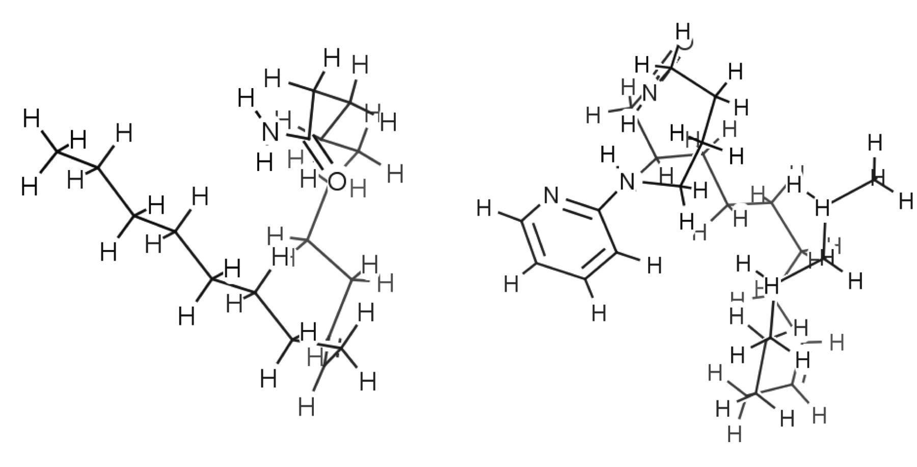
Most stable structures of compound 1 (left) and 18 (right).
Anti-HIV activity. In contrast to popular anti-HIV agents (dextran sulfate, curdlan sulfate, azidothymidine, 2’,3’-dideoxycytidine with SI=1935, 6028, 10403 and 1916, respectively), none of 18 oleoylamides protected the cells from the cytopathic effect of HIV infection (SIs<1) (Table III). Based on these data, the QASR analysis was focused on the cytotoxicity of oleoylamides.
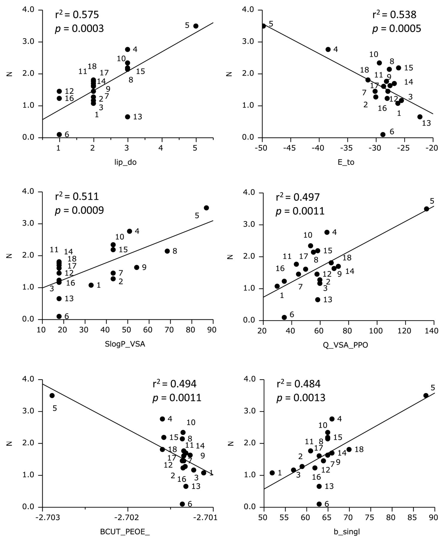
Computational analysis. All calculations of QSAR analysis of 18 oleoylamides were carried-out with MOE, using 330 descriptors including Hamiltonian (AM1. PM3 and MNDO). When the most stable structures were determined, all compounds exhibited compactly folded structures (Figure 2). QSAR analysis of cytotoxicity against normal cells demonstrated that 75 descriptors were correlated with N [mean pCC50 (i.e., −log CC50) for normal cells]. Scatter plots of the top six descriptors [lip_don (r2=0.575, p=0.0003), E_tor (r2=0.535, p=0.0005), SlogP_VSA0 (r2=0.511, p=0.0009), Q_VSA_PPOS (r2=0.497, p=0.0011), BCUT_PEOE_0 (r2=0.494, p =0.0011), b_single (r2=0.484, p=0.0013)] are shown in Figure 3. QSAR analysis of cytotoxicity against tumor cells demonstrated that 73 descriptors showed correlation with T (mean pCC50 for tumor cells). Scatter plots of the top six descriptors [PEOE_VSA-0 (r2=0.482, p=0.0014), PM3_HOMO (r2=0.477, p=0.0015), PM3_IP (r2=0.477, p=0.0015), SlogP_VSA0 (r2=0.450, p=0.0023), lip_don (r2=0.450, p=0.0023), PEOE_VSA+2 (r2=0.437, p=0.0028)] are shown in Figure 4. QSAR analysis of selective cytotoxicity demonstrated that nineteen descriptors showed correlation with T-N. Scatter plots of the top six descriptors [AM1_dipole (r2=0.387, p=0.0058), MNDO_dipole (r2=0.336, p=0.0117), PM3_HOMO (r2=0.334, p=0.0121), PM3_IP (r2=0.334, p=0.0121), dipole (r2=0.301, p=0.0184), logP(o/w) (r2=0.287, p=0.0220)] are shown in Figure 5. The tumor-specificity seems to increase when molecular polarization decreases and the hydrophobicity increases.
Anti-HIV activity of 18 oleoylamides and chemotherapeutic agents. Each value represents the mean of triplicate determinations.

Determination of correlation between chemical descriptors and mean cytotoxicity of oleoylamides against normal cells (defined as N). lip_don, The number of OH and NH atoms; E_tor, torsion (proper and improper) potential energy; SlogP_VSA0, sum of vi such that Li ≤−0.4; Q_VSA_PPOS, total positive polar van der Waals surface area; BCUT_PEOE_0, the smallest eigenvalues in the BCUT_PEOE; b_single, number of single bonds.

Determination of correlation between chemical descriptors and mean cytotoxicity of oleoylamides against tumor cells (defined as T). PEOE_VSA-0, Descriptors that depend on the partial charge of each atom of a chemical structure require calculation of those partial charges; PM3_HOMO, the energy (eV) of the highest occupied molecular orbital calculated using the PM3 Hamiltonian (MOPAC); PM3_IP, the ionization potential (kcal/mol) calculated using the PM3 Hamiltonian (MOPAC); SlogP_VSA0, sum of vi such that Li ≤−0.4; lip_don, the number of OH and NH atoms; PEOE_VSA+2, sum of vi where qi is in the range from 0.10 to 0.15.
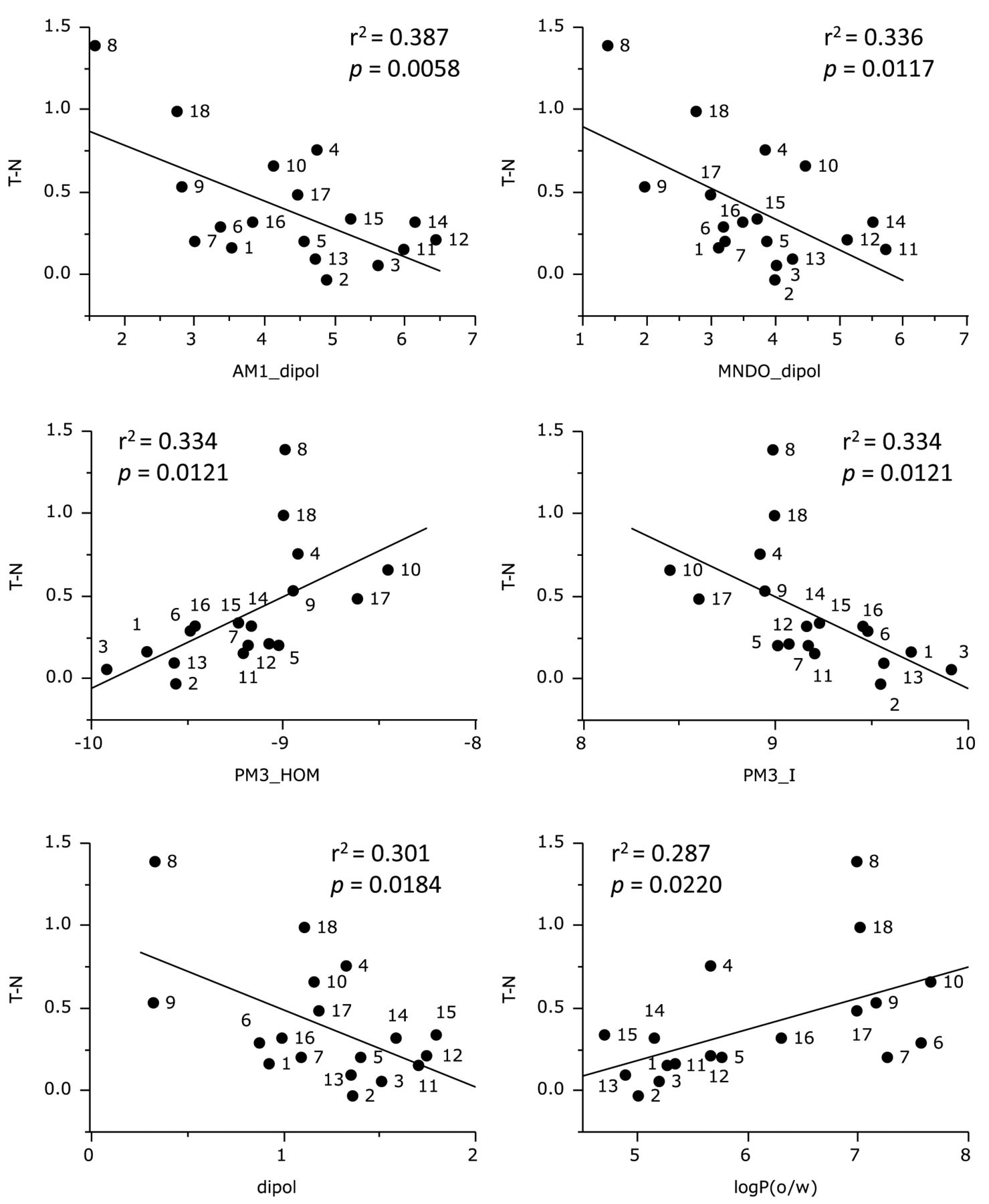
Determination of correlation between chemical descriptors and tumor specificity of oleoylamides (defined as T-N). AM1_dipole, The dipole moment calculated using the AM1 Hamiltonian (MOPAC); MNDO_dipole, the dipole moment calculated using the MNDO Hamiltonian (MOPAC); PM3_HOMO, the energy (eV) of the highest occupied molecular orbital calculated using the PM3 Hamiltonian (MOPAC); PM3_IP, the ionization potential (kcal/mol) calculated using the PM3 Hamiltonian (MOPAC); dipole, dipole moment calculated from the partial charges of the molecule; logP(o/w), log of the octanol/water partition coefficient.
We searched the combination sets that are useful for estimating cytotoxicity and tumor-selectivity using the stepwise method with leave-one-out cross verification. We constructed a good estimation model for N, T and T-N, using two or three descriptors each (Figure 6), producing the following equation:

Discussion
The present study demonstrated for the first time that 18 oleoylamides exhibit moderate to potent TS (Tables I, II) and no detectable anti-HIV activity (Table III). When cytotoxicity was compared between OSCC and normal mesenchymal cell (HGF, HPLF, HPC), compound [8] that has a catechol moiety had the highest TS (TS=15.5), exceeding that of 5-FU (TS=3.5), but of one order less than that of doxorubicin (TS=256) (Table I). Since compound [8] was identical with N-oleoyldopamine isolated from mammalian brain as endogenous capsaicin-like lipid (26), this compound should affect the cellular function in humans. It has been reported that N-oleoyl taurine increased the number of cells in the sub-G1 population, suggesting the induction of apoptosis, and a lower number of cells in the S-phase of the cell cycle (11). It remains to be investigated whether compound [8] has similar actions against OSCC cell lines.
When cytotoxicity was compared between OSCC and normal epithelial cells (HOK and HGPE), compound [18] which has a (2-pyridyl)amino group had the highest TS value (31.0 and 23.1, respectively), exceeding that of doxorubicin (0.3, and 0.033, respectively) and 5-FU (3.8 and 0.045, respectively) (Table II). With HOK and HGPE, oleoylamides [4], [5], [8] and [18] had much higher PSE values (638-14409 and 96-6661, respectively) compared to doxorubicin (516 and 55, respectively) and 5-FU (1.4 and 0.017, respectively), possibly due to highly cytotoxic properties of these chemotherapeutic agents. The high sensitivity of HOK and HGPE to doxorubicin may be due to the accelerated growth enforced by specially manufactured media for these epithelial cells.
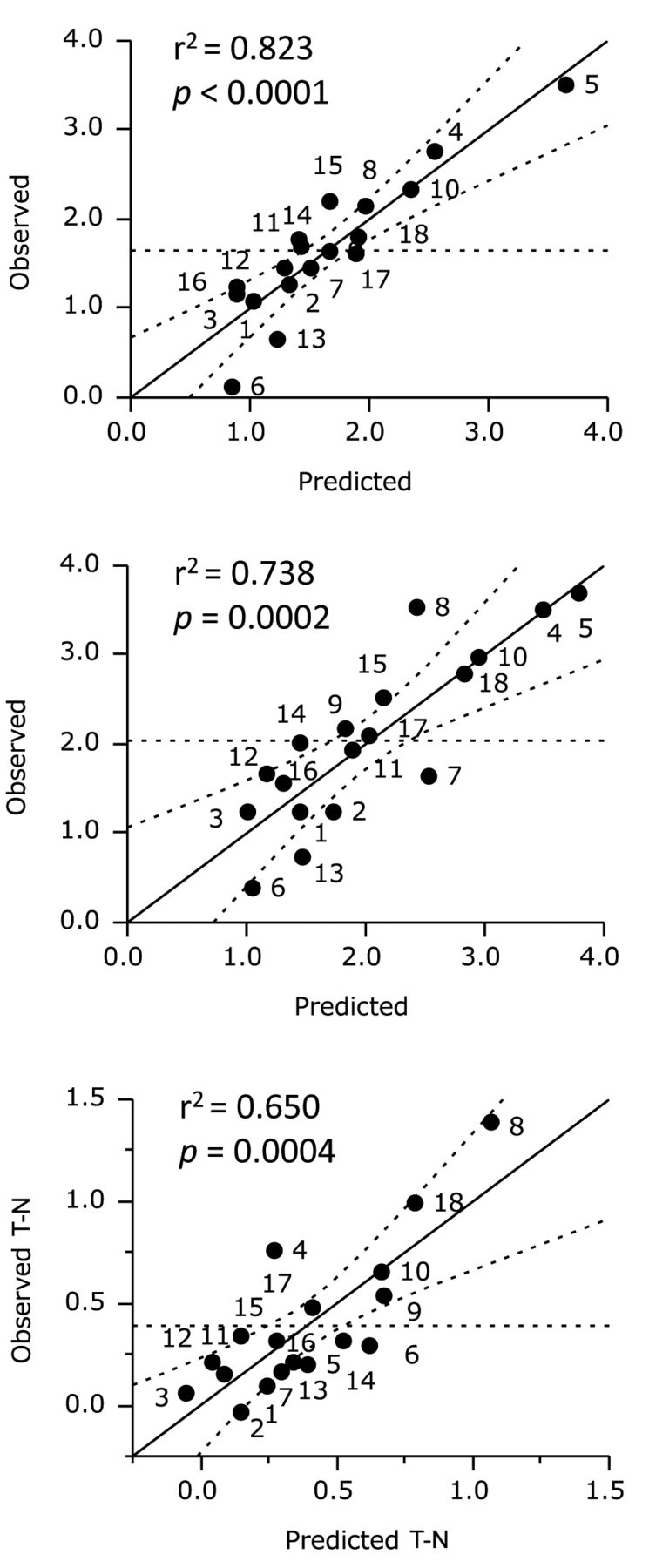
Multiple regression models for the estimation of tumor-specificity (T-N). T-N=32.1(±4.39)vsurf_R+121(±17)E_oop − 46.1 (±6.2); n=16, R2=0.870, Q2=0.821, s=0.145.
QASR analysis demonstrated that cytotoxicity against normal cells correlated well with six descriptors (lip_don, E_tor, SlogP_VSA0, Q_VSA_PPOS, BCUT_PEOE_0, b_single) that reflect the number of OH and NH groups, torsion potential energy, surface area and hydrophobicity, orbital electronegativity, and number of single bonds. (Figure 3). Cytotoxicity against tumor cells correlated well with six descriptors (PEOE_VSA-0, PM3_HOMO, PM3_IP, SlogP_VSA0, lip_don, PEOE_VSA+2) that reflec the partial charge of each atom, HOMO energy, ionization potential, surface area and hydrophobicity, and the number of OH and NH groups (Figure 4). TS correlated well with six descriptors [AM1_dipole, MNDO_dipole, PM3_HOMO, PM3_IP, dipole, logP(o/w)] that reflect the dipole moment, HOMO energy, ionization potential, and hydrophobicity (Figure 5). By searching various combination sets, we constructed a good estimation model for N, T and T-N, using two or three descriptors.
As far as we know, there is no study that has focused on the anti-viral activity of oleoylamides. This study demonstrated that none of the 18 oleoylamides protected the cells from the cytopathic effect of HIV infection. Further study is required to test whether these compounds have any other antiviral activity.
In conclusion, the present study suggests that multivariate statistics with chemical descriptors for molecular shape and flatness may be useful for evaluation of tumor-specificity of oleoylamides.
Acknowledgements
This work was partially supported by KAKENHI from the Japan Society for the Promotion of Science (JSPS) (15K08111). The annual license of the statistical software, JMP Pro, was supported by the grant-in-aid of the oncology specialists promotion program by the Ministry of Education, Culture, Sports, Science and Technology, Japan.
Footnotes
Conflicts of Interest
The Authors wish to confirm that there are no known conflicts of interest associated with this publication and there was no significant financial support for this work that could have influenced its outcome.
- Received June 24, 2015.
- Revision received July 16, 2015.
- Accepted July 20, 2015.
- Copyright© 2015 International Institute of Anticancer Research (Dr. John G. Delinassios), All rights reserved
References
In this issue


Jump to section
Related Articles
Cited By...
- Photodynamic Therapy With Resveratrol and an Nd:YAG Laser for Enterococcus faecalis Elimination
- Photodynamic Therapy with Pyoktanin Blue and Diode Laser for Elimination of Enterococcus faecalis
- In Vitro Antitumor Activity of Alkylaminoguaiazulenes
- Re-evaluation of Culture Condition of PC12 and SH-SY5Y Cells Based on Growth Rate and Amino Acid Consumption
- Search for New Type of Anticancer Drugs with High Tumor Specificity and Less Keratinocyte Toxicity
- Evaluation of Biological Activity of Mastic Extracts Based on Chemotherapeutic Indices
- Induction of Apoptosis in Human Oral Keratinocyte by Doxorubicin
- Quantitative Structure-Cytotoxicity Relationship of Chalcones
- Quantitative Structure-cytotoxicity Relationship of 3-Benzylidenechromanones
- Enhancement of Cytotoxicity of Three Apoptosis-inducing Agents Against Human Oral Squamous Cell Carcinoma Cell Line by Benzoxazinotropone