


Abstract
Background: Lenalidomide is an IMiD® immunomodulatory drug, which may warrant evaluation in urothelial carcinoma (UC). Materials and Methods: The in vitro and in vivo activity of lenalidomide was evaluated in human and murine UC cell lines. Tumors were evaluated by immunohistochemistry for (CD31), cleaved caspase-3 (CC3) and CD3+/CD20+ lymphocyte infiltration. Cereblon, a molecular target of lenalidomide was analyzed by immunohistochemistry. Results: Significant pro-apoptotic activity, and reduction of cell viability was seen at low micromolar concentrations of lenalidomide against indolent human RT4 UC cells in vitro. Cereblon expression was quantitatively lower in sensitive RT4 cells compared to resistant 5637 cells. In RT4 xenografts, lenalidomide significantly reduced tumor size and CD31 expression, and increased expression of CC3 (p<0.05). Cereblon expression increased in lenalidomide-treated RT4 xenografts (p<0.05). Conclusion: Lenalidomide demonstrated preclinical activity against superficially-invasive low-grade UC cells attributable to direct tumor cell apoptosis and anti-angiogenic activity. Clinical trials are warranted in patients with indolent UC.
The medical management of urothelial carcinoma (UC) has not enjoyed significant advances for over two decades. Although the initial response rates with cisplatin-based frontline combination chemotherapy are high (40% to 70%), the 5-year overall survival (OS) is only 4% to 20% (1-3). Multiple agents have demonstrated limited activity in the second-line setting (4-14). The modest success of intravesical instillation of Bacillus Calmette–Guérin (BCG) and interferon in high-risk non-muscle-invasive bladder cancer suggests a role for immunotherapy. Furthermore, angiogenesis in general, and vascular endothelial growth factor in particular, appear to be reasonable therapeutic targets. Interest in the activity of the combination of bevacizumab with cisplatin-based chemotherapy was a basis for the ongoing phase III trial investigating the impact of bevacizumab as a component of first-line therapy for advanced UC (15).
Lenalidomide is a US Food and Drug Administration-approved therapy for multiple myeloma and myelodysplastic syndromes associated with deletion 5q cytogenetic abnormality (16-19). Additionally, lenalidomide is feasible and demonstrates biological and antitumor activity in selected patients with advanced solid tumors (20, 21). Cereblon, the substrate receptor of the cullin ring E3 ubiquitin ligase 4 complex (CRL4CRBN), is a target of lenalidomide (22). Lenalidomide exerts its antiproliferative activity in multiple myeloma and also stimulates T-cells in a cereblon-dependent manner (22). The role of cereblon in anti-angiogenic activity and in cell autonomous effects in solid tumors is not yet known. Given the excellent tolerability to lenalidomide and likely relevance of such biological activity in UC, a rationale can be proposed to investigate the preclinical activity of lenalidomide in murine urothelial cancer models. Promising preclinical activity in an identifiable molecular subset of these carcinomas is relevant to the rational clinical development of this agent.
Materials and Methods
Cell lines and reagents. The in vitro anti-tumor activity of lenalidomide was evaluated in six human (5637, TCC-SUP, RT4, RT112, Hu456, T24) and one murine (MB49) cell lines. The 5637, TCC-SUP, RT4 and T24 cells were obtained from the American Type Culture Collection (Manassas, VA, USA). The RT112 and Hu456 cell lines were a gift provided by the laboratory of Dr. Ian C. Summerhayes (Beth Israel Deaconess Medical Center, Boston, MA, USA). 5637 was cultured as a monolayer in RPMI-1640 (Invitrogen, Grand Island, NY, USA) supplemented with 2.5 g/l of glucose, 2 mmol of L-glutamine, 1 mmol of sodium pyruvate, 10 mmol of HEPES, 10% fetal bovine serum, and 1% penicillin-streptomycin. TCC-SUP was cultured as a monolayer in Eagle's Minimum Essential Medium (Invitrogen) with 10% fetal bovine serum, and 1% penicillin-streptomycin. The RT4 and T24 cells were cultured as a monolayer in McCoy's 5a medium (Invitrogen) with 10% fetal bovine serum, and 1% penicillin-streptomycin. The RT112 and Hu456 cells were cultured in RPMI-1640 with 10% fetal bovine serum and 1% penicillin/streptomycin. MB49 cells were grown in grown in Dulbecco's modified Eagle's medium supplemented with 10% fetal bovine serum, 1% penicillin/streptomycin. All cells were grown in a humidified incubator with 5% carbon dioxide, at 37°C.
Lenalidomide for in vitro and in vivo use. Lenalidomide was provided by Celgene® (Summit, NJ, USA) for in vitro use. It was dissolved in Dimethyl sulfoxide (DMSO) and kept as a stock solution at a concentration of 100 mM. The solution was aliquoted and kept at −80°C until use. Frequent freeze-thawing was avoided. For in vivo use, lenalidomide was dissolved in water and this solution was kept at −20°C for up to seven days.
Cell viability (total cellular metabolism). For cell functional viability and activity assays, cells were plated in 96-well plates and total cellular metabolic activity was determined by the 3-(4,5-dimethylthiazol-2-yl)-2,5-diphenyltetrazolium bromide (MTT) colorimetric assay (Sigma-Aldrich, St. Louis, MO, USA) in replicates. Medium containing specified concentrations of the different chemotherapeutic agents was added to the wells and the cells were incubated for 72 h. Following incubation, MTT was added to each well, and the cells were incubated for an additional 4 h at 37°C. MTT reacts with mitochondrial metabolites to form a formazan salt resulting in a colorimetric change. Differences in total cellular metabolism were detected at a wavelength of 570 nm using a Fluostar Optima plate reader (BMG Labtech, Inc., Cary, NC, USA). Results were expressed as a relative percentage of total cellular metabolism compared with that of untreated controls (controls expressed as 100%). The MTT assays were also performed on cells exposed for an extended duration to lenalidomide at 1 μmol or DMSO alone (control). In these experiments, the medium was changed daily.
In addition to the MTT assay, the cytotoxic effect of lenalidomide was assessed by measuring the number of viable cells using crystal violet. Ninety-six-well plates were seeded at 150 cells per well. After 24 h, cells were incubated in the absence or presence of 1 μM lenalidomide and the medium was changed daily, for 10-14 days. The medium was then aspirated and the cells were fixed with 10% buffered formalin. After 10 min, the 10% buffered formalin was removed and the cells were stained with 1% crystal violet in 70% ethanol for 3 min, followed by washing with tap water and allowing to air dry. After air-drying, the retained crystal violet was dissolved in 100 μl of 30% methanol/10% acetic acid; absorbance was read at 590 nm.
Apoptosis assay. Fluorescein isothiocyanate (FITC) annexin V is used to quantitatively determine the percentage of cells within a population that are actively undergoing apoptosis. Cells were treated with 2.2 μM lenalidomide for 72 h, changing medium daily for three days. Cells were collected and stained with annexin-V and propidium iodide (PI) and analyzed by Fluorescence Activated Cell Sorting.
Administration of agents to mice bearing subcutaneous tumor grafts. All experiments involving animals were carried out under approved protocol (AN-4104) granted by the Institutional Animal Care and Use Committee of Baylor College of Medicine between February 2012 to December 2014. Six to eight week old female nude mice (The Jackson Laboratory, Bar Harbor, Maine, USA) were given ectopic tumors on their right flank by injection of 5×10⁶ cells mixed 1:1 with matrigel, s.c. (100 μl/mouse). After approximately three weeks of growth, tumor size was measured and mice were assigned to either the control or treatment group. Two cohorts of 10 nu/nu mice per group each bearing subcutaneous RT4 tumors were assigned to groups in a way that that the baseline average and standard deviation of tumor sizes were equal. Mice were given either placebo or 10 mg/kg lenalidomide once daily for five days a week for four weeks by oral gavage in a volume of 0.1 ml. In a similar separate experiment, two cohorts of 15 C57BL/6 mice (The Jackson Laboratory, Bar Harbor, Maine, USA), each bearing subcutaneous MB49-Luc25 (luciferase labeled MB49 cells) murine cell tumors were administered placebo or 10 mg/kg lenalidomide once daily for five days a week for four weeks. Tumor dimensions in all mice were measured using a caliper and the tumor volume estimated using the formula: m12 × m2 × 0.5236, where m1 is the length of the short axis and m2 is the length of the long axis of the tumor.
Immunohistochemistry of murine xenografts and cell-lines. Tumors removed from the mice after sacrifice were fixed in formaldehyde and embedded in paraffin. Expression of cluster of differentiation-31 (CD31), cleaved-caspase-3 (CC3), cereblon and CD3/CD20 (only in MB49-Luc25 tumors in C57BL/6 mice) were analyzed in five randomly selected murine xenografts from each group by immunohistochemistry (IHC) of histological sections. Appropriate positive and negative controls were also stained. Four high-power fields (×400) were examined per tumor. Each image was quantified for immunoreactivity using a 0 to 3+ scoring system for both the intensity of staining and the percentage of positively stained cells by usingImage-Pro Plus Software (Media Cybernetics, Rockville, MD, USA). Cells were considered positive for cereblon if they displayed at least 2+ expression, whereas for CC3 and CD31, cells with any degree of expression were considered positive.
Statistical analysis. The significance of differences was determined by Student's t-test using JMP 5.1.2 statistical software package (SAS Institute, Cary, NC, USA). p-Values were considered significant by Student's t-test if p≤0.05.
Results
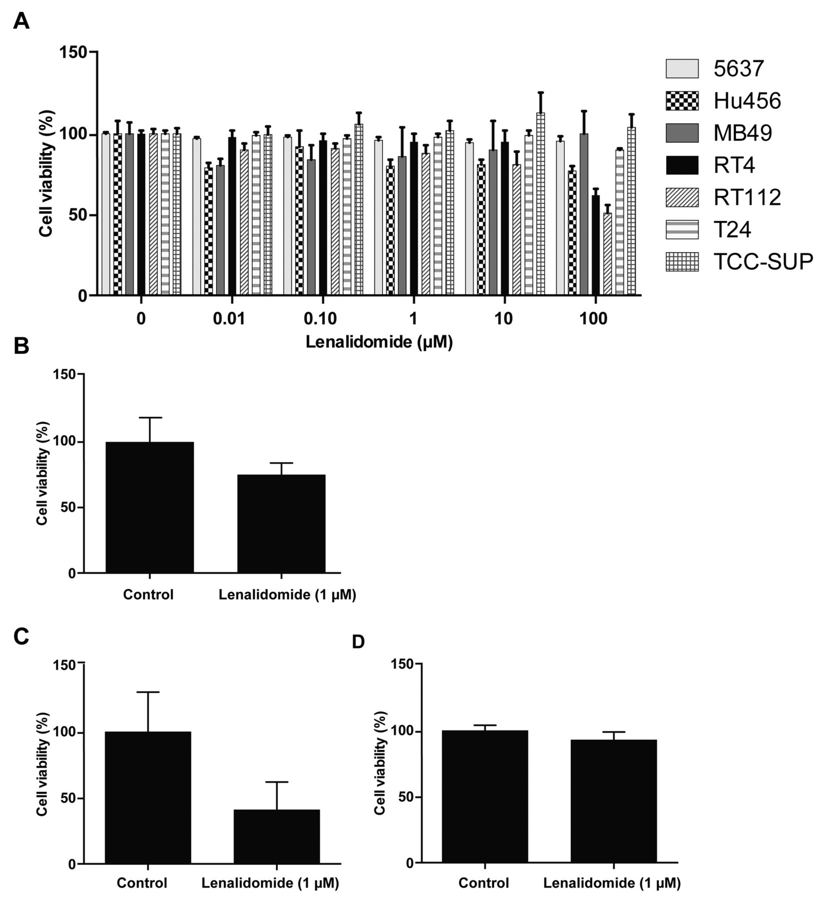
Differential sensitivity to lenalidomide in bladder cancer cell lines. Exposure for 72 h with daily change of medium demonstrated marginal or no alterations in cellular metabolism against all cells at physiologically-attainable low micromolar concentrations of lenalidomide (1-10 μM), as measured by the MTT assay (Figure 1A). High concentrations of lenalidomide (100 μM) demonstrated a modest activity in RT4 and RT112 cells. Modest activity (p<0.01) was also observed after 14 days of exposure to 1 μM lenalidomide of RT4 cells with daily change of medium (Figure 1B), but not in the other cell lines (data not shown). To further evaluate the differential sensitivity of bladder cancer cell lines to lenalidomide, a sensitive line (RT4) and a resistant line (5637) were treated with 1 μM lenalidomide for 14 days as described above, and a crystal violet assay was performed to assess cellular viability. The lenalidomide treatment led to 42% inhibition of RT4 cells compared to the vehicle-treated control (p<0.0001; Figure 1C). No cytotoxic effects were seen in 5637 cells following the same treatment (Figure 1D). Induction of apoptosis was observed in the sensitive RT4 cell line, which exhibited significant apoptotic induction after three days' exposure to as little as 2.2 μM lenalidomide, as measured by annexin-V staining (Figure 2A). The assay of the 5637 cells showed no apoptosis under similar conditions (data not shown).

In vitro activity of lenalidomide in urothelial carcinoma cell lines 3-(4,5-dimethyl-2-thiazolyl)-2,5-diphenyl-2H-tetrazolium bromide assay after exposure to 1-10 μM lenalidomide for 72 h with daily change of medium demonstrated marginal to no alterations of metabolism in all cell lines, while 100 μM lenalidomide demonstrated modest activity in RT4 and RT112 cells (A). Exposure to lenalidomide at 1 μM of RT4 cells with daily change of medium for 14 days showed modest activity (B). Crystal violet assay to assess cellular viability after treatment with 1 μM lenalidomide for 14 days showed 42% inhibition in RT4 cells compared to vehicle control (p<0.0001; C) but no effects in 5637 cells (D).

Pro-apoptotic and in vivo activity of lenalidomide in RT4 cells. Induction of apoptosis (annexin-V staining) was observed in sensitive RT4 cells after three days' exposure to 2.2 μM lenalidomide (A). After four weeks of lenalidomide treatment, growth of subcutaneous RT4 xenografts was significantly inhibited compared to those from mice treated with vehicle-only (p<0.05; B).
Lenalidomide has significant activity against RT4 bladder tumors in in vivo xenografts. Four weeks of receiving the lenalidomide treatment significantly inhibited tumor growth in nude mice bearing RT4 xenografts compared to mice receiving vehicle-only (p<0.05; Figure 2B). In contrast, lenalidomide at 10 mg/kg daily orally for five days a week for up to four weeks did not reduce the size of MB49-Luc25 subcutaneous tumors in syngeneic immunocompetent C57BL/6 mice compared to control mice (data not shown).
Immunohistochemistry of xenografts shows modulation of cereblon, of angiogenesis and of apoptosis by lenalidomide. Cereblon expression was quantitatively lower in untreated RT4 cells compared to 5637 cells (data not shown, p>0.05). The cereblon expression in tumors of mice following lenalidomide therapy was increased (p<0.05) compared to controls, with 60% of tumors showing high (3+) cereblon staining (Figure 3A). Mice treated with lenalidomide exhibited lower expression of the vascular marker, CD31 (p<0.05), suggesting that lenalidomide may have an anti-angiogenic effect (Figure 3B). Furthermore, RT4 tumors from mice treated with lenalidomide exhibited a significant increase of the apoptotic marker CC3 (p<0.05; Figure 3C). The IHC of MB49-Luc25 syngeneic tumors in immunocompetent C57BL/6 mice showed significant reduction in CD31 (p<0.05) on treatment with lenalidomide compared to controls, but no statistical differences for CC3 or CD3/CD20+ lymphocytes (data not shown).
Discussion
In our preclinical study, lenalidomide treatment demonstrated selective preclinical activity against superficially-invasive low-grade RT4 human UC cells, attributable to direct tumor cell apoptosis and anti-angiogenic activity. Indeed, an immuno-competent model showed anti-angiogenic activity, but no retardation of growth in vivo. This may be a result of the absence of tumor cell pro-apoptotic activity against the MB49 cell line. The immunocompetent animal model also did not demonstrate immunomodulation measured as CD3/CD20+ lymphocytes. Thus, direct inhibition may be the most important mechanism conferring antitumor activity in the studied models.
However, the anti-tumor activity of lenalidomide appeared to be cytostatic and was most evident after long-term continuous exposure. In addition, the sensitive RT4 cells quantitatively demonstrated lower cereblon expression, although the trend was not statistically significant for these experiments. Even more intriguingly, lenalidomide treatment statistically significantly appeared to increase cereblon expression in RT4 xenografts. These data complement other data from a group of investigators demonstrating that lenalidomide augments the response to BCG in a different in vivo immunocompetent mouse model of non-muscle-invasive bladder cancer (MBT-2 cell line implanted in C3H mice) (23). Lenalidomide reduced both tumor burden and microvessel density in that prior study.
Thalidomide, and now lenalidomide, has been identified to bind to cereblon and form an E3 ubiquitin ligase complex which does inhibit ubiquitin ligase activity (24). Thereafter, the anti-tumor activity of lenalidomide in multiple myeloma was also found to be associated with higher cereblon expression, particularly in the context of IMiD drug resistance (25-28). Unlike the association of high cereblon expression with activity of lenalidomide in multiple myeloma, our study detected a signal of better activity with lower cereblon expression, which increased after lenalidomide exposure in vivo. Therefore, the role, if any, of cereblon as a predictive biomarker needs further investigation with regard to the potential for UC to be susceptible to lenalidomide. In this context, the role of targeting cereblon in the activity of lenalidomide in the 5q myelodysplastic syndrome remains unclear. It remains important that the pleiotropic properties of lenalidomide encompassing anti-angiogenic and immunomodulating activity be addressed.

Immunohistochemistry of RT4 xenografts treated with lenalidomide vs. controls Cereblon expression following lenalidomide therapy in RT4 xenografts was increased (p<0.05) compared to that of controls (A). Xenografts treated with lenalidomide exhibited lower expression of cluster of differentiation (CD)-31 (p<0.05, B) and a significant increase of cleaved-caspase 3 (CC3) (p<0.05; C).
A limitation of our study is partly a consequence of the limited resources available for further interrogating the mechanism of activity in UC. Following the MTT assay in six human UC cell lines, we chose to concentrate resources by following signals from the MTT assay. To this end, we further investigated one potentially sensitive (RT4) and one resistant (5637) human cell line in vitro by flow cytometry for apoptosis and by crystal violet assay for viability. We assessed one murine cell line in a syngeneic murine model (MB49-Luc25) and one human cell line (Hu456) in a xenograft model. The cell line used for the xenograft model was selected for sensitivity to lenalidomide in vitro.
Lenalidomide appeared to be cytostatic in vivo in the xenograft and did not reduce tumor burden. Moreover, lenalidomide appeared to demonstrate no anti-tumor activity in the murine syngeneic immunocompetent model, despite reducing angiogenesis. Moreover, B- and T-lymphocyte infiltration in the tumor was not amplified by lenalidomide. Notably, the cell line used for the syngeneic immunocompetent model was not sensitive to lenalidomide in vitro. These data suggest that the direct anti-tumor cell activity of lenalidomide may be more critical than the effects of lenalidomide on the microenvironment, the immune system and on angiogenesis in the context of UC.
To summarize, we demonstrated that lenalidomide increased apoptosis and reduced tumor growth, in some cases, in superficially-invasive indolent bladder cancer cells at low micromolar concentrations which are clinically-relevant and attainable in humans. Given that persistent or progressive non-muscle-invasive bladder cancer following intravesical BCG has no effective therapy, lenalidomide may warrant consideration for this indication in a trial also addressing potentially predictive biomarkers, such as cereblon. Additional mechanistic studies to define the circumstances when immunomodulation or anti-angiogenic activity are relevant, as opposed to a direct pro-apoptotic effect, will help define the profile of urothelial tumors that could be targeted with lenalidomide. The NCT01373294 trial is evaluating the combination of intra-vesical BCG and oral lenalidomide together in patients with Ta high-grade, T1 high-grade or carcinoma in situ following prior BCG treatment. In this trial, patients receive daily oral lenalidomide for seven months, overlapping three courses of BCG, at the start of treatment, at three months, and at six months, with the hypothesis that the immune response to BCG can be augmented. In addition to lenalidomide, other IMiD® immunomodulatory drugs, particularly pomalidomide, may also warrant evaluation in preclinical models to address cytotoxic, cytostatic, immune, and anti-angiogenic activities that could be relevant to a clinical impact on UC or other types of solid cancers.
Acknowledgements
We thank Dr. Hiroshi Handa from Integrated Research Institute, Tokyo Institute of Technology, Yokohama 226-8503, Japan, for kindly supplying antibodies to cereblon and Mayer B. Fishman, MD, Ph.D. for critical reading of the manuscript.
Footnotes
-
* Presented in part as a poster at the Genitourinary Cancer Symposium, Orlando, Florida in February 2013.
-
Conflicts of Interest
Weiguo Jian, Jonathan M. Levitt, Seth P. Lerner, Guru Sonpavde received Research support to their institution by Celgene.
-
Funding Sources
Celgene.
- Received January 29, 2014.
- Revision received May 6, 2014.
- Accepted May 7, 2014.
- Copyright© 2014 International Institute of Anticancer Research (Dr. John G. Delinassios), All rights reserved
References
In this issue


Jump to section
Related Articles
Cited By...
- No citing articles found.