


Abstract
Background: Biotin is an essential micronutrient and its levels are high in rapidly proliferating cells such as cancer cells. We hypothesized that the synthesis of poly (amido)amine (PAMAM) dendrimers with biotin molecules might contribute to enhanced cancer cell-specific uptake. Materials and Methods: PAMAM dendrimers were biotinylated using sulfo-NHS-LC-biotin and structural characterization was performed using 1H NMR and matrix assisted laser desorption ionization — time-of-flight. The effect of generation and the mechanism of cellular uptake of biotin-PAMAMG4 in ovarian cancer (OVCAR-3) and human embryonic kidney (HEK 293T) cells was determined by fluorescent microscopy and flow cytometry. Results: The cellular uptake of Biotin-PAMAM was significantly higher in the OVCAR-3 cells as compared to the HEK 293T cells (p<0.05). While the presence of either free biotin or colchicine significantly reduced the extent of uptake of biotin-PAMAMG4 at lower concentration (0.1 μM). The results demonstrated that the biotinylated-PAMAM was internalized by biotin receptor-mediated endocytosis and charge-mediated adsorptive endocytosis. The cytotoxicity of biotinylated-PAMAMG4 in the HEK 293T cells was comparable to that of the parent PAMAM dendrimers. Conclusion: Biotinylated PAMAM dendrimers show potential as nanocarriers in targeted drug delivery.
Ovarian cancer is considered as a chemosensitive gynecological malignancy and it is the fourth most frequent cause of cancer death among women (1-3). Even after clinically and surgically defined complete response to chemotherapy, the majority of women experience recurrence of the cancer within two years (4). These latter tumors generally do not respond to chemotherapy due to the development of drug resistance and spread into the peritoneal cavity resulting in the development of ascites. Therefore, it has been suggested that the delivery of chemotherapy beyond the ‘standard dosage regimen’ (standard six courses) might be capable of killing the sensitive micro tumors that were less vulnerable to cytotoxic agents during the primary treatment (5). Targeted delivery of high doses of chemotherapeutics using cancer cell-specific ligands may be an attractive alternative for the successful treatment of these resistant tumors (6-11). One such important ligand investigated for targeted drug delivery in recent years is biotin.
Biotin is an essential micronutrient for normal cellular functions (e.g. fatty acid biosynthesis, gluconeogenesis), growth and development. Humans and other mammals cannot synthesize biotin and thus must obtain it from exogenous sources via intestinal absorption. Rapidly dividing cells such as cancer cells have a voracious appetite for certain vitamins including biotin, vitamin B12 and folate and biotin levels have been found to be significantly higher in some cancer cells compared to normal tissue (12). Interestingly, tumor cell lines including ovarian, colorectal etc., which overexpress receptors involved in folate or vitamin B12 uptake also showed overexpression of biotin receptors (13). Accordingly, several research groups have tried different biotinylated chemotherapeutic agents for cancer cell-specific drug delivery (14-16). For example, biotinylated poly(ethyleneglycol) conjugates of camptothecin have been shown to be more cytotoxic and enhanced the ability to induce apoptosis by activation of the caspase-dependent cell death signaling pathway simultaneously suppressing antiapoptotic cellular defense and were effective in multidrug resistant ovarian carcinoma cells 30 times more compared with free camptothecin, opening up a new application of biotinylation in overcoming resistance to chemotherapy (16). Nevertheless, biotin-drug conjugates suffer from two major limitations, only small doses of drug can be delivered as only one drug molecule can be attached per ligand and the ligand-drug conjugates are very small and so will be excreted by the kidney and may be reabsorbed in the proximal tubules, leading to undesirable accumulation in the kidney. Biotinylated polymeric carrier systems may be a suitable alternative to overcome these limitations. Several research groups have documented the extravasation of nanoparticles of less than 200 nm from the tumor vascular endothelium and their trapping because of lack of lymphatic drainage in the tumor tissue, together termed the enhanced permeability and retention (EPR) effect. Krueger et al. have covalently grafted biotin to nanodiamond without any loss of biological activity and suggested various biological, medical and drug delivery applications of these nanobiotin devices (17). Biotinylated poly(ethyleneglycol)-block-poly(N- sopropylacrylamide-co-N-hydroxymethylacrylamide), a thermo responsive micelle showed increased uptake in tumor cells (18). Biotinylated poly(lactic acid)-poly(ethyleneglycol) [PLA-PEG] nanoparticles significantly increased the cytotoxicity of paclitaxel when compared to Taxol® and nanoparticles without biotin as targeting ligand (19).
Dendrimers are synthetic nanometer range polymers with unique attributes such as tailorable surface moieties, low polydispersity index and biocompatibility and have been widely used in tumor drug targeting (8, 20-24). Poly(amido)amine dendrimers (PAMAM) have been widely investigated in tumor specific targeting of anticancer drugs and macromolecules (25). Several different tumor-specific ligands covalently conjugated to dendrimers have been used to target LHRH receptors, αVβ3 integrin, epidermal growth factor receptor and folate receptors (26-29). Thus, dendrimers are considered to be versatile nanocarriers for ligand based tumor specific targeting and are an attractive approach for obtaining high drug concentration at the tumor site (30-32). Different generations of PAMAM dendrimers are commercially available, at a relatively low cost and the toxicity of the dendrimers increases with increase in generation number (33).
In the present study, biotin-PAMAM dendrimer conjugates were synthesized and characterized and the effect of different generations of the dendrimers on cellular uptake by ovarian cancer cells was investigated. Cancer cell specificity of the biotin-conjugated dendrimers was evaluated quantitatively using flow cytometry by comparing the uptake between human embryonic kidney (HEK 293T) and ovarian cancer (OVCAR-3) cells. The biocompatibility of biotin-dendrimer conjugates was also assessed by determining the cytotoxicity to HEK 293T cells.
Materials and Methods
PAMAM-NH2 dendrimers with an ethylenediamine core and amine surface groups of different generations were obtained from Dendritic Nanotechnologies Inc., Mount Pleasant, MI, USA, and 3-[4,5-dimethylthiazol-2-yl]-2,5-diphenyl tetrazolium bromide (MTT), D2O, dihydrobenzoic acid (DHB), tetramethyl silane (TMS), 4-(2-hydroxyethyl)-1-piperazineethanesulfonic acid (HEPES), fluorescein isothiocyanate (FITC), biotin, colchicine and dimethylsufoxide (DMSO) were purchased from Sigma-Aldrich (Saint-Louis, MO, USA). Sulfo-NHS-LC-biotin (Mr 556) and the avidin/hydroxyazobenzene-2-carboxylic acid (HABA) reagent was from Pierce Chemical Co., Rockford, IL, USA. Trifluoroacetic acid (TFA), ethylene diamine tetra acetic acid (EDTA), phosphate buffered saline (PBS, pH 7.4), acetone and acetonitrile (both High Performance Liquid Chromatography grade) and cell culture materials were purchased from Fisher Scientific (Pittsburgh, PA, USA). Hank's balanced salt solution (HBSS) with calcium and magnesium (without phenol red) was purchased from Cellgro (Herndon, VA, USA). Dialysis bags were purchased from Spectrum Laboratories, Rancho Dominguez, CA, USA.
Biotinylation of dendrimers. The biotin-conjugated PAMAM dendrimers were prepared as reported previously (34). A schematic representation of the method is shown in Figure 1. At room temperature, 20 mg each of generations G1, G2, G3 and G4 PAMAM dendrimers (20 mg in 2 mL of 0.1 M phosphate buffer (pH 9.0) were added to sulfo-NHS-LC-biotin at a molar ratio of 1:20 for G1 and G2 generations, and 1:30 for G3 and G4 generations and stirred for 2 h. The mixture was then dialyzed against deionized water to remove unconjugated biotin. The solution was then lyophilized (Virtis Benchtop K series, Virtis, Gardiner, NY, USA) to yield a white product.
Hydroxyazobenzene-2-carboxylic acid (HABA) assay. The degree of biotinylation of the biotinylated PAMAM dendrimers was determined by HABA assay. Briefly, the avidin/HABA reagent was prepared according to the manufacturer's instructions by adding 10 mg of avidin and 600 μL of HABA solution (24.2 mg in 9.9 mL of water and 0.1 mL of 1N NaOH) to 19.4 mL phosphate buffered saline (PBS pH 7.4). Then 100 μL of biotinylated dendrimer dissolved in deionized water was added to a 900 μL of the avidin-HABA solution, and the absorbance was measured at 500 nm using a SpectraMax M2 microplate reader (Molecular Devices, Sunnyvale, CA, USA). A schematic illustration of the assay is shown in Figure 2.
NMR and MALDI analysis. Proton nuclear magnetic resonance spectra (1H NMR) (chemical shift in ppm with respect to TMS set at zero) of the biotinylated conjugates were obtained on a 400 MHz Bruker AMX-400 spectrometer (Bruker, Rheinstetten, Germany), with samples dissolved in deuterium oxide (D2O) at a concentration of 5 mg/mL. To confirm the degree of biotinylation, mass spectral analysis of the dendrimers was performed using a Bruker MALDI-TOF (matrix assisted laser desorption/ionization-time-of flight). The dendrimer samples were prepared by diluting to a 10 mg/mL solution in methanol. A 10 mg/mL solution of 2,5 dihydroxybenzoic acid in 0.1 M TFA was used as the matrix solution. The dendrimer and matrix solution were mixed at a ratio of 1:5 and spotted onto the steel grid and dried for the analysis.
Fluorescence labeling of biotinylated PAMAM dendrimers. Briefly, 10mg of the biotinylated dendrimers (G1-G4) were dissolved in PBS (pH 7.4) in a round bottomed flask. A 5 mg/mL solution of FITC in acetone was prepared and added to the dendrimer solution at a molar ratio of 1:1. The mixture was kept under stirring for 4 h at room temperature. The mixture was dialyzed against 4 L of deionized water and lyophilized. Fluorescently labeled dendrimer solution was purified by Sephacryl S-300 column chromatography with acetonitrile:Tris buffer (70:30) as the elution buffer. The elution fractions corresponding to the dendrimer size were collected, dialyzed against deionized water at 4°C, lyophilized and stored at 4°C for further studies. The stability of the FITC-labeled dendrimers was studied at 4°C and 37°C (for 5 days) by determining the free FITC in the samples using Agilent 1100 high pressure liquid chromatography with fluorescence detector (Agilent Technologies, Santa Clara, CA, USA). Phosphate buffered saline (PBS pH 7.4): acetonitrile (80:20) was used as the mobile phase. No second peak representing free FITC (retention time of 5.5 min) was found in the samples indicating that the labeled dendrimers were stable for 5 days. The extent of FITC conjugation was obtained using a SpectraMax M2 microplate reader with 485 nm as the excitation wavelength and 525 nm as the emission correlating the absorbance with a FITC calibration curve.
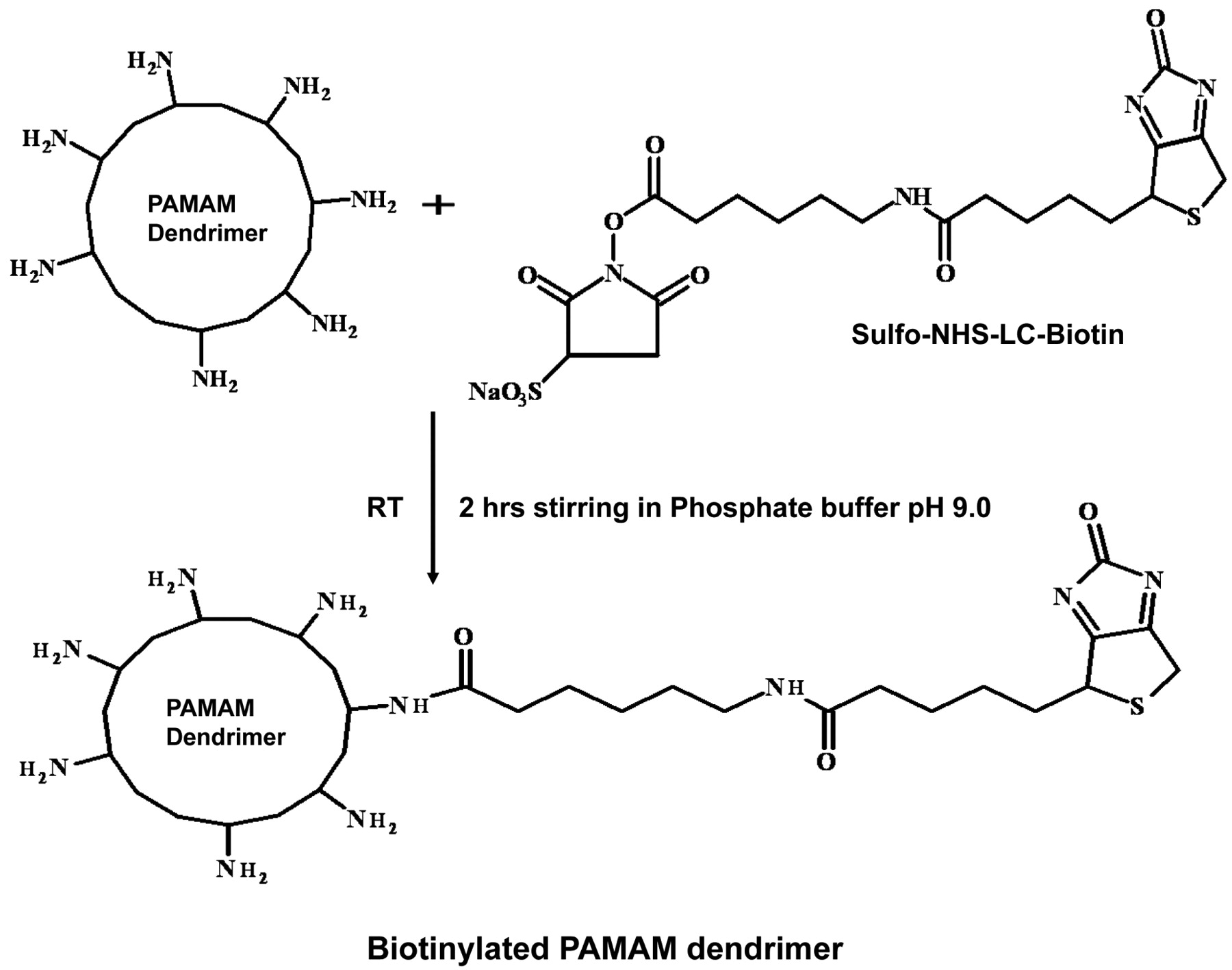
Conjugation of biotin to the amine terminals of the PAMAM dendrimers using NHS-LC-biotin [N-hydroxysuccinimidyl-6-(biotinamido) hexanoate] as the biotinylation reagent (Note: The number of surface amine groups increases with the increase in generation number of the PAMAM dendrimers. The figure does not represent any particular generation of dendrimers). RT: room temperature.
Cell culture. Human ovarian cancer (OVCAR-3) and human embryonic kidney (HEK 293T) cells were obtained from American type cell culture (ATCC, Manassas, VA, USA). The cells were grown in an atmosphere of 5% CO2 and 95% relative humidity using RPMI 1640 Medium (pH 7.4) supplemented with 10% fetal bovine serum and 1% penicillin-streptomycin solution (Cellgro, Mediatech Inc., Herndon, VA, USA).
Fluorescence microscopy. The OVCAR-3 and HEK 293T cells were seeded at a density of 1×10⁵ cells per well in 12 well plates (Multiwell, Becton Dickinson Labware, Franklin lakes, NJ, USA) 24 h before the experiment. The cells were then incubated with the FITC-labeled biotinylated PAMAM dendrimers (G1-G4) at 10 μg/mL concentration and maintained at 37°C with 5% CO2 for 2, 4, 6, 8 or 24 h. At the end of the incubation period, the medium was removed, the cells were washed with 3×1 mL of PBS and incubated in 1 mL of PBS. Fluorescence micrographs of the cells were taken using an Olympus IX70 inverted-microscope (Olympus America Inc., Center valley, PA, USA) equipped with a filter for FITC.
In vitro cytotoxicity assay. In vitro cytotoxicity of the dendrimers was evaluated by MTT assay (35). HEK 293T cells were seeded at a density of 2×10⁴ cells per well in a 96 well plate. Following 24 h incubation, different concentrations (10-200 μg/mL) of the parent PAMAM dendrimers and biotinylated dendrimers (G1-G4) were added. After a 4 h incubation period, 50 μL of 1:10 diluted MTT stock solution (5 mg/mL) was added and the cells were incubated for 4 h. The medium was removed, 150 μL of DMSO was added to dissolve the MTT crystals and the optical density was read using a SpectraMax M2 microplate reader (Molecular devices, Sunnyvale, CA) with 590 nm as excitation wavelength and 650 nm as the background. The viability of the cells exposed to the dendrimers was expressed as a percentage of the viability of the cells grown in the absence of dendrimers.
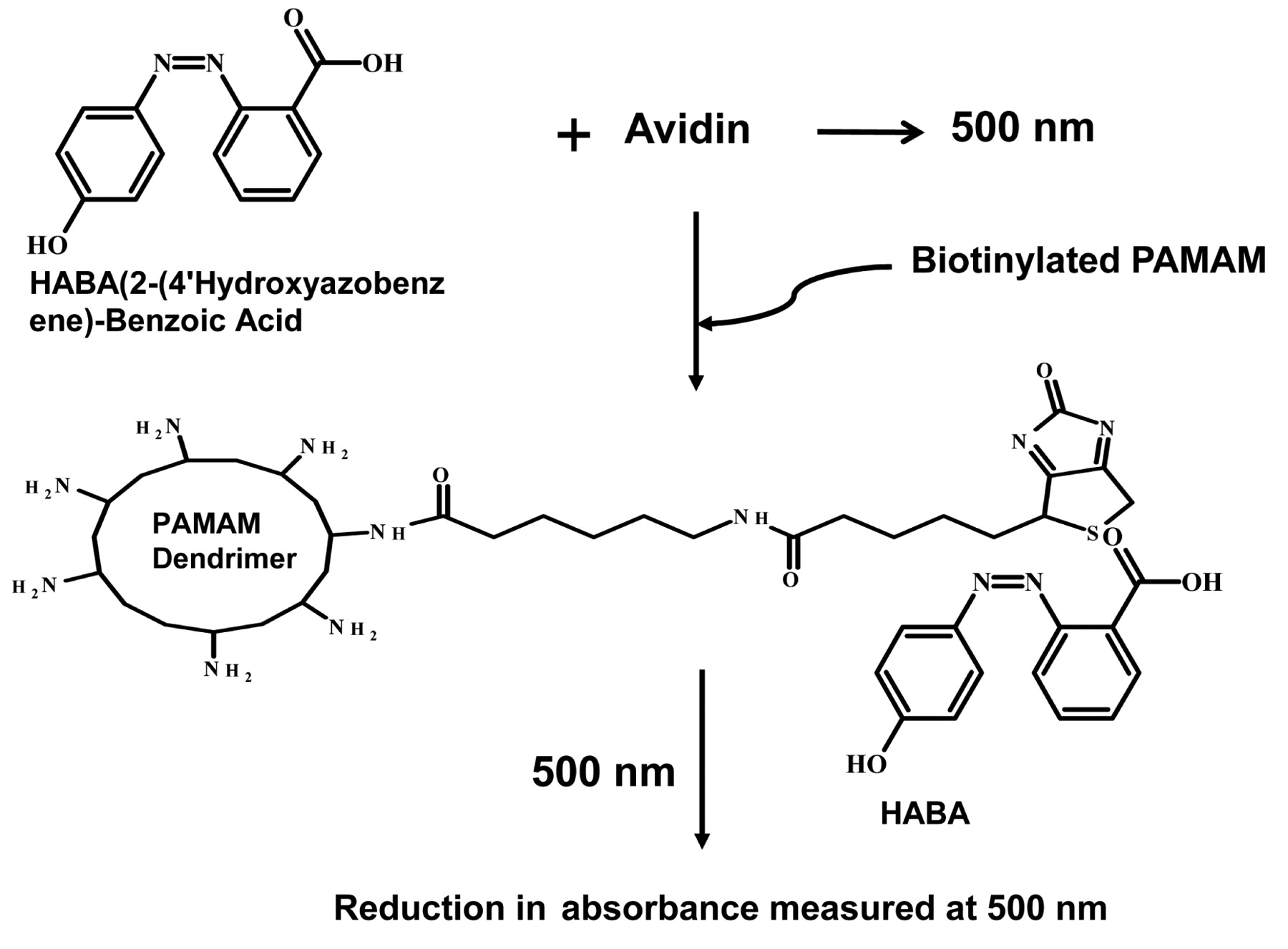
Hydroxyazobenzene-2-carboxylic acid (HABA) assay procedure. Avidin/HABA reagent was prepared as per manufacturer's instructions, 100 μL of biotinylated PAMAM dendrimer solution was added to 900 μL of the avidin/HABA reagent solution and the absorbance was measured at 500 nm using a microplate reader.
Cellular uptake study. To investigate if biotinylated dendrimers are internalized by receptor mediated endocytosis, dendrimer cellular uptake studies were performed as follows: 1×10⁵ OVCAR-3 or HEK-293T cells per well were seeded in 12 well plates 24 h prior to the experiment. The cells were incubated with 10 μM colchicine or 1 mM biotin for 30 min prior to treatment with different concentrations of biotinylated dendrimers in separate experiments and incubated at 37°C for 1 h. The cells were washed three times with PBS pH 7.4, trypsinized and suspended in 200 μL of PBS containing 2% fetal bovine serum. The intensity of fluorescence in the cells was detected and corrected for background fluorescence of the control cells using a fluorescence-activated cell sorting (FACS) machine with argon ion laser of 488 nm (Beckton Dickinson Inc. CA, USA) and the data was processed with Cell-Quest Pro software.
Statistical analysis. The statistical analysis was performed using two-way ANOVA with general linear model (Graph Pad InStat 3.05 version software (San Diego, CA, USA) followed by multiple comparison and p<0.05 was considered statistically significant.
Results
The biotinylation reaction resulted in a high yield (>85%) and the final product was a fluffy white fibrous solid. The 1H NMR data revealed the presence of biotin ring juncture protons at δ=4.4 and 4.6 ppm which were conspicuously absent in the parent PAMAM dendrimers (36), however, the other characteristic peaks of the dendrimer (2.6-3.6 ppm) were observed in both the parent and biotinylated PAMAM dendrimers (Figure 3). The extent of biotinylation of the various generations of dendrimers was quantified using the HABA assay. About 2, 3, 6 and 7 biotin molecules were conjugated to G1, G2, G3 and G4 PAMAM dendrimers respectively. To further confirm the conjugation and extent of biotinylation, the biotin-PAMAM dendrimers were analyzed using MALDI-TOF spectroscopy. For the G1, G2 and G3 generations, the extent of biotinylation as calculated by MALDI-TOF was the same as that of the HABA assay i.e. 2, 3 and 6 biotin molecules respectively (data not shown). However, MALDI-TOF depicted 27 biotin molecules conjugated to PAMAMG4 dendrimers. It may be inferred from the above results that the HABA assay was not accurate in measuring the higher degree of biotinylation of dendrimers (Figure 4).

1H NMR of PAMAMG4 (A) and biotinylated PAMAMG4 (B) with D2O as solvent and 5 mg/mL concentration.
The effect of the PAMAM generation on the cellular uptake of the dendrimers was determined by FITC labeling. Fluorescent micrographs revealed that the increase in generation from G1 to G4 resulted in an increase in the uptake (Figure 5). It was also found that the intensity of fluorescence in the OVCAR-3 cells was greater than that of in HEK 293T cells. As evident from the fluorescent micrographs data, the generation 4 dendrimers showed the highest intensity of fluorescence.
PAMAM dendrimers of generation 4 were chosen to study the in vitro cellular uptake as measured by flow cytometry. As shown in Figure 6, the uptake of the FITC-conjugated biotinylated PAMAMG4 dendrimers was 12% higher in the OVCAR-3 cells when compared with the HEK 293T cells.
The cellular uptake of FITC-conjugated biotinylated PAMAMG4 dendrimers was also measured in the presence of biotin (1 mM) or an endocytosis inhibitor, colchicine (10 μM), in separate experiments. While the cellular uptake of the dendrimers in the absence of biotin and colchicine was 90.23±4.2%, the presence of biotin and colchicine significantly reduced the uptake of the dendrimers to 78.24±0.19 and 54.12±1.48% respectively (p<0.001). However, at higher concentrations of the dendrimer (1 μM and 10 μM), the presence of biotin or colchicine did not affect the extent of uptake of the biotinylated dendrimers (Figure 7).
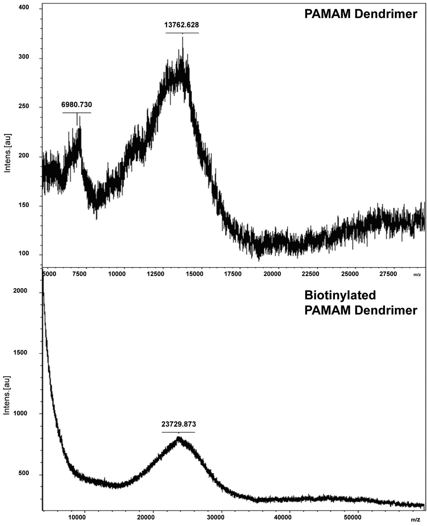
MALDI-TOF spectra of PAMAMG4 and biotinylated PAMAMG4.
The cytotoxicity of the biotinylated PAMAM dendrimers to the HEK 293T cells was assessed using the MTT assay. At 200 μg/mL, cell viability decreased from 67.51±1.5% to 40.02±1.75% as the generation number increased from G1 to G4 (p<0.05). The in vitro cytotoxicity of the biotinylated dendrimers was comparable to that of the parent PAMAM dendrimers and they were found to be safe up to a concentration of 100 μg/mL. Above 100 μg/mL, the biotinylated PAMAM dendrimers were slightly more toxic than the parent PAMAM dendrimers (Figure 8).

In vitro cellular uptake studies in OVCAR-3 and HEK 293T cells. Cells in fluorescent mode (FM) (left panels) and bright mode (BM) (right panels). A1 and A2, B1 and B2: Biotin-PAMAMG1 A3 and A4, B3 and B4: Biotin-PAMAMG2; A5 and A6, B5 and B6: Biotin-PAMAMG3 and A7 and A8, B7 and B8: Biotin-PAMAMG4.
Discussion
The biotinylation method used was rapid with a yield always >85% and involved the use of the water soluble biotinylation reagent, sulfo-NHS-LC-biotin. This is a derivative of D-biotin containing a spacer arm off the valeric acid side-chain with N-hydroxysuccinimide (NHS) ester end group. The most important advantage of using this reagent compared to simple biotin is that, the 6-aminocaproic acid spacer provides greater length between the covalently modified molecule and the bicyclic biotin rings. This would result in better binding potential for the biotin receptors expressed over the cancer cells. Moreover, the extended length would also reduce the steric hindrance due to the biotin rings for further attachments.
The NMR spectroscopy of the biotinylated dendrimers revealed the presence of two protons of biotin. Though the HABA assay has been used for quantifying the extent of biotinylation very extensively, the present results from MALDI-TOF suggest that its accuracy is limited to lower generations of dendrimers as the HABA assay suffers from steric hindrance in determining the extent of biotinylation in highly biotinylated molecules (12, 34, 38).
Unlike many ligands (such as hormones), vitamins are internalized by receptor mediated endocytosis and biotin is taken up by a sodium dependent multivitamin transport system (SMVT) (37). It is likely that a carrier system such as dendrimers conjugated to vitamins would be taken by the same transport system. The PAMAMG4 dendrimers have 64 terminal amine groups and it was assumed that the synthesis of a dendrimer that has surplus of spatially oriented biotin molecules might contribute to enhanced uptake by the ovarian cancer cells. FITC was chosen as a marker because it involves a simple reaction to bind to biotinylated dendrimers and it also exerts a strong fluorescence for efficient identification of the molecule when internalized in cells (39, 40). The microscopic studies revealed greater internalization of biotinylated dendrimers when compared with the parent PAMAM dendrimers validating the hypothesis that biotinylated dendrimers can be used as carriers for rapid internalization via biotin/SMVT receptors. Moreover, the biotinylated dendrimers from G1 to G4 showed increasing uptake which was consistent with previous data by Kelly and co-workers in a Caco-2 cell permeability model (41), and may be attributed to the greater cationic surface charge density that enabled increased interaction with receptors on the cell membrane. The data was encouraging in that the increased paracellular permeability of the higher generation PAMAM dendrimers combined with enhanced cancer cellular internalization mediated by biotin makes this carrier system very effective in cancer cell targeting and permeation across the tumor tissue.

In vitro cellular uptake of PAMAMG4 in OVCAR-3 and HEK-293T cells measured by flow cytometry. Left: FACS analysis and right: percent cellular uptake of FITC-labeled PAMAM and biotinylated PAMAM dendrimers.
As expected, as shown by flow cytometry the biotinylated dendrimers were internalized 12% more in the OVCAR-3 than in the HEK 293T cells. The increase could be attributed to the fact that ovarian cancer cells which grow rapidly require more biotin as it is required for the expression of oncogenes and cell proliferation. It was also reported previously that, of all vitamin (folic acid, vitamin B12 and biotin)-conjugated chemotherapeutic agents biotin targeted conjugate appears to possess the greatest antitumor activity and showed significantly greater reduction in tumor load which was attributed to the overexpression of biotin receptors in tumor cells (13).

Mechanism of cellular uptake of FITC-conjugated biotinylated dendrimers. OVCAR-3 cells incubated with 1 mM biotin or 10 μM colchicine 30 min prior to treatment with different concentrations of biotinylated PAMAMG4 dendrimer. The intensity of fluorescence in the cells was detected using FACS.
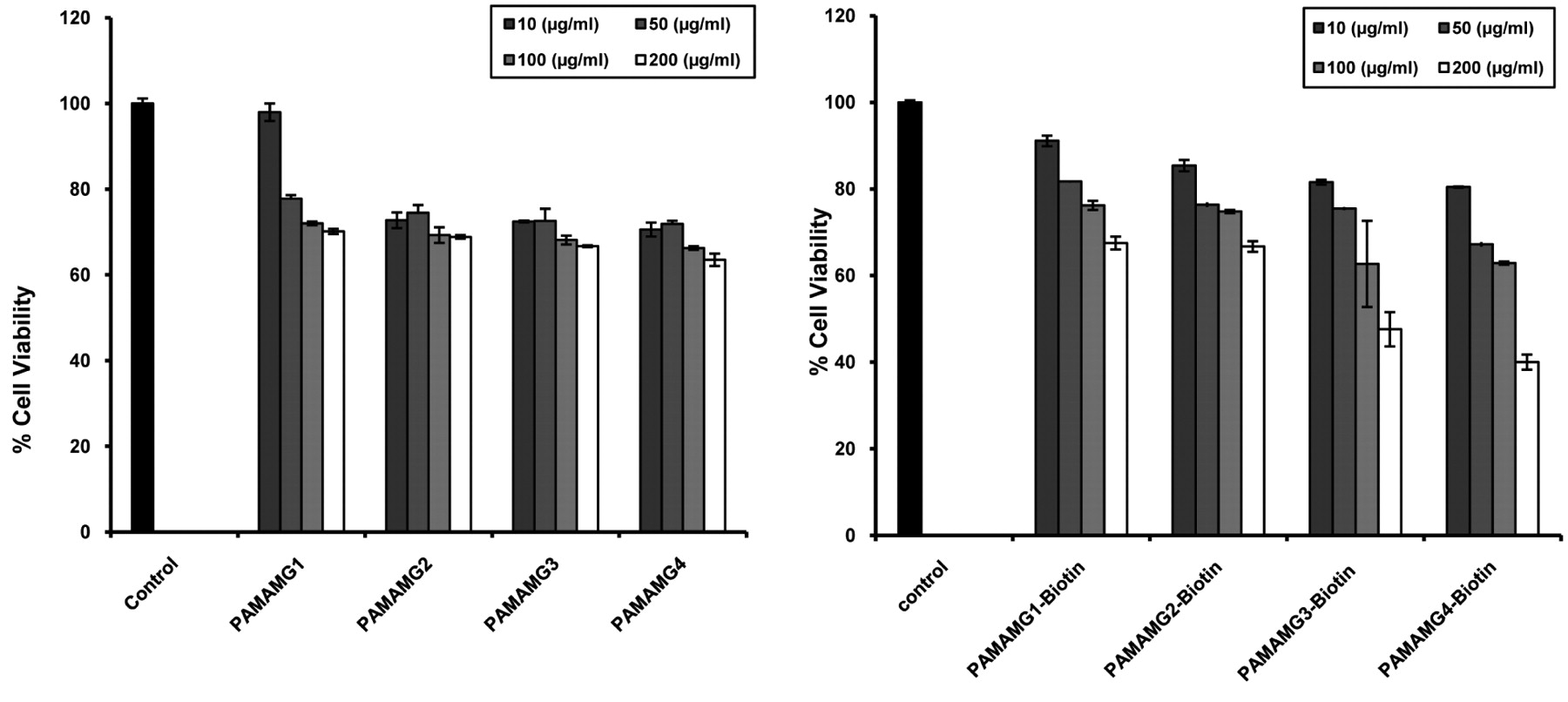
Effect of generation number of PAMAM and biotinylated-PAMAM dendrimers on the viability of HEK293T cells at different concentrations.
Although biotin has been used as a ligand for cancer cell targeting, the mechanism of cellular uptake of biotin-conjugated nanoparticles has not been investigated. One of the main objectives of the study was to determine the role of biotin receptors/SMVT system in the cellular internalization of biotin-conjugated PAMAM dendrimers. Understanding the mechanism of biotinylated PAMAM dendrimers uptake may help in improving the targeting efficiency of this carrier system. Cationic dendrimers such as PAMAM-NH2 are commonly reported to undergo cellular internalization by endocytosis mechanisms (41). It is therefore, reasonable to expect that even biotinylated dendrimers would be internalized by endocytosis. Interestingly, the presence of colchicine (10 μM) significantly reduced the cellular uptake of biotinylated dendrimers at lower concentrations (p<0.01), indicating that endocytosis was involved in their uptake. However, to determine if biotin receptor mediated endocytosis has a role for this carrier system, the cancer cells were incubated with free biotin followed by incubation with the biotinylated dendrimers. Whilst the uptake of dendrimers at the lower concentration (0.1 μM) was significantly reduced in the presence of biotin, the presence of biotin did not inhibit their uptake at higher concentrations. Thus, the involvement of both adsorptive endocytosis and biotin-receptor mediated endocytosis in the cancer cellular uptake of the biotinylated dendrimers was suggested. The charge mediated adsorptive endocytosis appeared to be the predominant mechanism as the dendrimer uptake was not inhibited in the presence of biotin when used at higher concentrations.
In order to serve as efficient universal scaffolds for drug delivery as well as targeting, the carrier system needs to be biocompatible. It may be assumed that carrier systems with rapid cellular internalization may be cytotoxic in vivo. The MTT assay, results suggested that the toxicity of the biotinylated dendrimers was comparable to that of the native counterparts and increased with increase in generation number. As cellular uptake and cytotoxicity of the biotinylated dendrimers increased with generation number, it may be suggested that higher uptake was the reason behind the cytotoxicity of the biotinylated PAMAM dendrimers.
In conclusion, biotinylated dendrimers might be exciting nano carriers which could be used both for antibody-based pretargeting and also SMVT receptor mediated targeting. Our future studies will include the use of these nano scaffolds for an antibody-based pretargeting strategy in a murine ovarian cancer model.
Acknowledgements
This work was supported by a grant from the National Institutes of Health (NIH R15 CA121980-01) to SP. The authors also thank Andrew Ramirez for his technical help in the preparation of the manuscript.
- Received March 2, 2009.
- Revision received June 2, 2009.
- Accepted June 12, 2009.
- Copyright© 2009 International Institute of Anticancer Research (Dr. John G. Delinassios), All rights reserved


